EasySep™小鼠Pan-ILC富集试剂盒

免疫磁阴性选择16分钟细胞分离

产品号 #(选择产品)

产品号 #19875_C

免疫磁阴性选择16分钟细胞分离

产品优势

  • 快速,易于使用和无列
  • 分离的细胞不受影响
  • 促进快速流动分类的ILCs

产品组分包括

  • EasySep™小鼠泛ilc富集试剂盒(目录#19875)
    • EasySep™小鼠Pan-ILC富集鸡尾酒,0.5 mL
    • EasySep™Streptavidin RapidSpheres™50001,1.0 mL
New look, same high quality and support! You may notice that your instrument or reagent packaging looks slightly different from images displayed on the website, or from previous orders. We are updating our look but rest assured, the products themselves and how you should use them have not changed. Learn more

概述

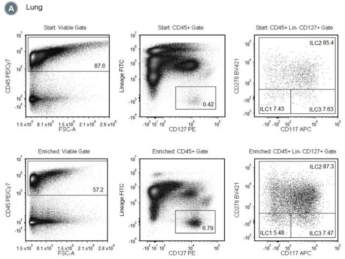
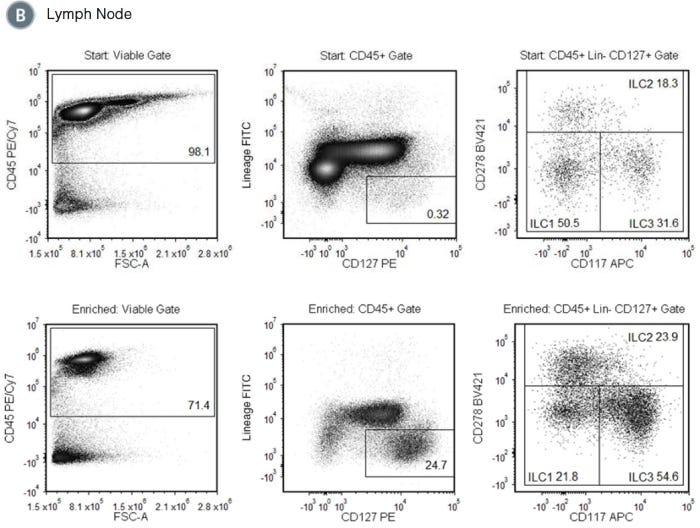
本品旨在通过负选择从小鼠组织单细胞悬液中富集1、2、3组先天淋巴样细胞(ILC1、ILC2和ilc3)。不需要的细胞是针对非ilcs和链亲和素包被磁颗粒(RapidSpheres™)的生物素化抗体靶向去除的。使用EasySep™磁铁分离标记细胞,不使用色谱柱。不需要的细胞留在管中,而需要的细胞只是被倒进一个新的管中。分离的细胞可立即用于下游应用,如流式细胞术和细胞分选。

Magnet Compatibility

• EasySep™ Magnet (Catalog #18000)
• EasyEights™ EasySep™ Magnet (Catalog #18103)
• EasyPlate™ (Catalog #18102)
 
Subtype
Cell Isolation Kits
 
Cell Type
Innate Lymphoid Cells
 
Species
Mouse
 
Sample Source
Bone Marrow, Lung, Other
 
Selection Method
Negative
 
Application
Cell Isolation
 
Brand
EasySep
 
Area of Interest
Immunology
 

Data Figures

Figure 1. Typical EasySep™ Mouse Pan-ILC Enrichment Profile

Starting with a naïve mouse lung single-cell suspension, the total ILC content (CD45+Lin-CD127+) of the enriched fraction typically ranges from 3.1 - 7.6%. In the above example, the percentages of ILCs in the start and final enriched fractions are 0.4% and 3.9% (or 0.4% and 6.8% of CD45+ cells), respectively. NOTE: The ILC content of the start fraction typically ranges from 0.3 - 0.6%.

Figure 2. Typical EasySep™ Mouse Pan-ILC Enrichment Profile from Mouse Lymph Node

Starting with a naïve mouse lymph node single-cell suspension, the total ILC content (CD45+Lin-CD127+) of the enriched fraction typically ranges from 21.1 - 45.2%. In the above example, the percentages of ILCs in the start and final enriched fractions are 0.3% and 17.6% (or 0.3% and 24.7% of CD45+ cells), respectively. NOTE: The ILC content of the start fraction typically ranges from 0.3 - 0.4%.

Protocols and Documentation

Find supporting information and directions for use in the Product Information Sheet or explore additional protocols below.

Document Type
Product Name
Catalog #
Lot #
Language
Catalog #
19875
Lot #
All
Language
English
Document Type
Safety Data Sheet 1
Catalog #
19875
Lot #
All
Language
English
Document Type
Safety Data Sheet 2
Catalog #
19875
Lot #
All
Language
English

Applications

This product is designed for use in the following research area(s) as part of the highlighted workflow stage(s). Explore these workflows to learn more about the other products we offer to support each research area.

Resources and Publications

Educational Materials (8)

Publications (3)

Mesenchymal Stem Cells Attenuate Asthmatic Inflammation and Airway Remodeling by Modulating Macrophages/Monocytes in the IL-13-Overexpressing Mouse Model. Y. Mo et al. Immune network 2022 oct

Abstract

Mesenchymal stem cells (MSCs) are attractive alternatives to conventional anti-asthmatic drugs for severe asthma. Mechanisms underlying the anti-asthmatic effects of MSCs have not yet been elucidated. This study evaluated the anti-asthmatic effects of intravenously administered MSCs, focusing on macrophages and monocytes. Seven-week-old transgenic (Tg) mice with lung-specific overexpression of IL-13 were used to simulate chronic asthma. MSCs were intravenously administered four days before sampling. We examined changes in immune cell subpopulations, gene expression, and histological phenotypes. IL-13 Tg mice exhibited diverse features of chronic asthma, including severe type 2 inflammation, airway fibrosis, and mucus metaplasia. Intravenous administration of MSCs attenuated these asthmatic features just four days after a single treatment. MSC treatment significantly reduced SiglecF-CD11c-CD11b+ monocyte-derived macrophages (MoMs) and inhibited the polarization of MoMs into M2 macrophages, especially M2a and M2c. Furthermore, MSCs downregulated the excessive accumulation of Ly6c- monocytes in the lungs. While an intravenous adoptive transfer of Ly6c- monocytes promoted the infiltration of MoM and Th2 inflammation, that of MSC-exposed Ly6c- monocytes did not. Ex vivo Ly6c- MoMs upregulated M2-related genes, which were reduced by MSC treatment. Molecules secreted by Ly6c- MoMs from IL-13 Tg mice lungs upregulated the expression of fibrosis-related genes in fibroblasts, which were also suppressed by MSC treatment. In conclusion, intravenously administered MSCs attenuate asthma phenotypes of chronic asthma by modulating macrophages. Identifying M2 macrophage subtypes revealed that exposure to MSCs transforms the phenotype and function of macrophages. We suggest that Ly6c- monocytes could be a therapeutic target for asthma management.
OASL1-Mediated Inhibition of Type I IFN Reduces Influenza A Infection-Induced Airway Inflammation by Regulating ILC2s. Y. Chang et al. Allergy, asthma & immunology research 2022 jan

Abstract

PURPOSE Three observations drove this study. First, 2'-5'-oligoadenylate synthetase-like protein (OASL) is a negative regulator of type I interferon (IFN). Second, type I IFN plays a central role during virus infections and the pathogenesis of various diseases, including asthma. Third, influenza A virus (IAV) causes non-eosinophilic asthma. To evaluate the potential relationships between OASL, type I IFN, and pulmonary innate immune cells in IAV-induced acute airway inflammation by using Oasl1-/- mice. METHODS Asthma was induced in wild-type (WT) and Oasl1-/- mice with IAV or ovalbumin (OVA). Airway hyperreactivity (AHR) and immune cell infiltration in the bronchoalveolar lavage (BAL) fluids were measured. The immune cells in the lungs were analyzed by flow cytometry. To investigate the ability of type I IFN to shape the response of lung type 2 innate lymphoid cells (ILC2s), IFN-$\alpha$ was treated intratracheally. Plasmacytoid dendritic cells (pDCs) sorted from bone marrow and ILC2s sorted from lungs of naive mice were co-cultured with/without interferon-alpha receptor subunit 1 (IFNAR-1)-blocking antibodies. RESULTS In the IAV-induced asthma model, Oasl1-/- mice developed greater AHR and immune cell infiltration in the BAL fluids than WT mice. This was not observed in OVA-induced asthma, a standard model of allergen-induced asthma. The lungs of infected Oasl1-/- mice also had elevated DC numbers and Ifna expression and depressed IAV-induced ILC2 responses, namely, proliferation and type 2 cytokine and amphiregulin production. Intratracheal administration of type I IFN in na{\{i}}ve mice suppressed lung ILC2 production of type 2 cytokines and amphiregulin. Co-culture of ILC2s with pDCs showed that pDCs inhibit the function of ILC2s by secreting type I IFN. CONCLUSIONS OASL1 may impede the IAV-induced acute airway inflammation that drives AHR by inhibiting IAV-induced type I IFN production from lung DCs thereby preserving the functions of lung ILC2s including their amphiregulin production."
Endothelin-A Receptor Antagonist Alleviates Allergic Airway Inflammation via the Inhibition of ILC2 Function. X. Zhang et al. Frontiers in immunology 2022

Abstract

Allergic airway inflammation is a universal airway disease that is driven by hyperresponsiveness to inhaled allergens. Group 2 innate lymphoid cells (ILC2s) produce copious amounts of type 2 cytokines, which lead to allergic airway inflammation. Here, we discovered that both peripheral blood of human and mouse lung ILC2s express the endothelin-A receptor (ETAR), and the expression level of ETAR was dramatically induced upon interleukin-33 (IL-33) treatment. Subsequently, both preventive and therapeutic effects of BQ123, an ETAR antagonist, on allergic airway inflammation were observed, which were associated with decreased proliferation and type 2 cytokine productions by ILC2s. Furthermore, ILC2s from BQ123 treatment were found to be functionally impaired in response to an interleukin IL-33 challenged. And BQ123 treatment also affected the phosphorylation level of the extracellular signal-regulated kinase (ERK), as well as the level of GATA binding protein 3 (GATA3) in activated ILC2s. Interestingly, after BQ123 treatment, both mouse and human ILC2s in vitro exhibited decreased function and downregulation of ERK signaling and GATA3 stability. These observations imply that ETAR is an important regulator of ILC2 function and may be involved in ILC2-driven pulmonary inflammation. Therefore, blocking ETAR may be a promising therapeutic strategy for allergic airway inflammation.

更多信息

更多信息
Species Mouse
Magnet Compatibility • EasySep™ Magnet (Catalog #18000) • EasyEights™ EasySep™ Magnet (Catalog #18103) • EasyPlate™ (Catalog #18102)
Sample Source Bone Marrow, Lung, Other
Selection Method Negative
标记抗体
PRODUCTS ARE FOR RESEARCH USE ONLY AND NOT INTENDED FOR HUMAN OR ANIMAL DIAGNOSTIC OR THERAPEUTIC USES UNLESS OTHERWISE STATED. FOR ADDITIONAL INFORMATION ON QUALITY AT STEMCELL, REFER TO WWW.STEMCELL.COM/COMPLIANCE.
版权 © 2025 STEMCELL Technologies 技术有限公司。保留所有权利。