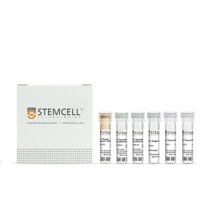
产品号 #19861_C
免疫磁珠负选不带标记的小鼠单核细胞
免疫磁珠负选不带标记的小鼠单核细胞
使用EasySep™小鼠单核细胞分选试剂盒,可通过负选,轻松高效地从小鼠骨髓、脾细胞、全血或其他单细胞悬液样本中分离高纯度的小鼠单核细胞。EasySep™技术结合单克隆抗体的特异性和无柱磁分选系统的简便性,已在发表的研究中广泛应用超过20年。
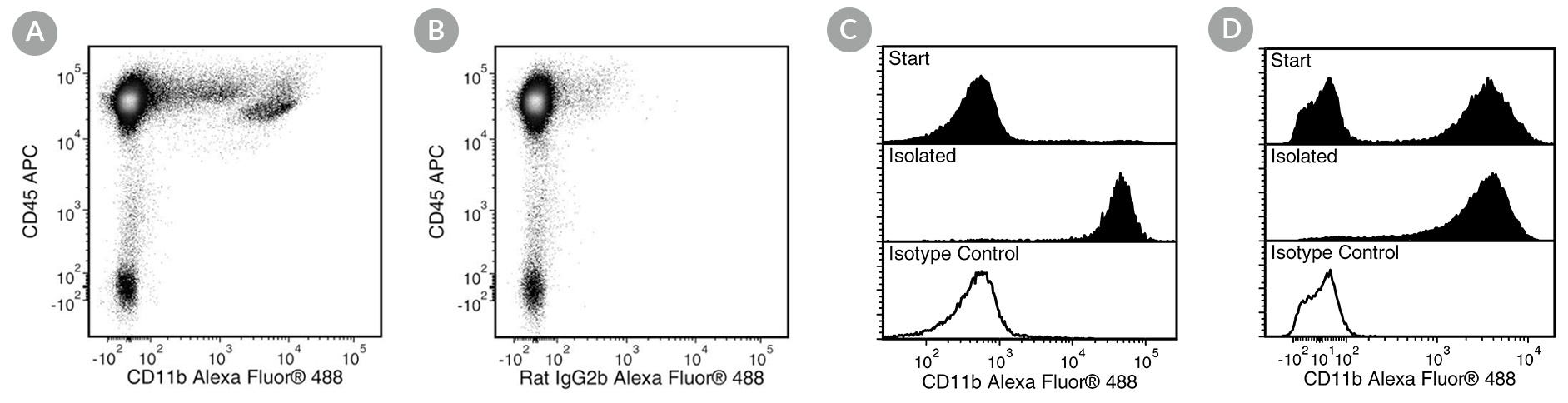
在该EasySep™负选流程中,非目的细胞通过抗体复合物与磁珠标记。以下非目的细胞将被靶向去除:粒细胞、T细胞、B细胞、NK细胞、造血祖细胞及红系细胞。使用EasySep™磁极吸附后,通过简单地将目的细胞倾倒或吸取至一个新的试管中,即可将被磁珠标记的细胞与不带标记的目的细胞分离开来。在短至15分钟的磁珠分选后,目的单核细胞可立即用于流式细胞术、细胞培养、基于细胞的实验等下游应用。
了解更多EasySep™免疫磁珠技术的工作原理,或者如何通过RoboSep™实现全自动化免疫磁珠细胞分选。探索更多为您的实验流程优化的产品,包括培养基、补充剂、抗体等。
磁体兼容性
• EasySep™ Magnet (Catalog #18000)
• “The Big Easy” EasySep™ Magnet (Catalog #18001)
• EasyEights™ EasySep™ Magnet (Catalog #18103)
• EasyPlate™ EasySep™ Magnet (Catalog #18102)
• RoboSep™-S (Catalog #21000)
亚型
细胞分选试剂盒
细胞类型
单核细胞
种属
小鼠
样本来源
Bone Marrow,Spleen,Whole Blood
筛选方法
Negative
应用
细胞分选
品牌
EasySep,RoboSep
研究领域
免疫
Find supporting information and directions for use in the Product Information Sheet or explore additional protocols below.
This product is designed for use in the following research area(s) as part of the highlighted workflow stage(s). Explore these workflows to learn more about the other products we offer to support each research area.
| Species | Mouse |
|---|---|
| Magnet Compatibility | • EasySep™ Magnet (Catalog #18000) • “The Big Easy” EasySep™ Magnet (Catalog #18001) • EasyEights™ EasySep™ Magnet (Catalog #18103) • EasyPlate™ EasySep™ Magnet (Catalog #18102) • RoboSep™-S (Catalog #21000) |
| Sample Source | Bone Marrow, Spleen, Whole Blood |
| Selection Method | Negative |
哺乳动物有核细胞手动计数试剂
计数板含有2个盖玻片
细胞分选缓冲液
全自动细胞分选仪器

亚美尼亚仓鼠单克隆IgG1抗体,抗小鼠CD3e

大鼠单克隆IgM抗体,抗小鼠CD49b(整合素α2)
大鼠单克隆IgG2a抗体,抗小鼠Ly-6G
大鼠单克隆IgG2a抗体,抗人、小鼠、猫CD45R(B220)

大鼠单克隆IgG2a抗体,抗小鼠F4/80

抗小鼠 NK1.1 (CD161) 的 (C3H x BALB/c) F1 杂交单克隆IgG2a抗体

抗人、小鼠、恒河猴CD11b的大鼠单克隆IgG2b抗体