EasySep™人浆细胞样DC富集试剂盒

免疫磁珠负选试剂盒

产品号 #(选择产品)

产品号 #19062_C

免疫磁珠负选试剂盒

产品优势

  • 操作简单、快捷,且无需分离柱
  • 纯度高达97%
  • 获得的活细胞无标记

产品组分包括

  • EasySep™人浆细胞样DC富集试剂盒(产品号 #19062)
    • EasySep™人浆细胞样DC富集抗体混合物组分A,2 x 1 mL
    • EasySep™人DC富集抗体混合物组分B,2 x 1 mL
    • EasySep™ D Magnetic Particles 磁珠,8 x 1 mL
    • 抗人CD32 (Fc gamma RII) 阻断剂,2 x 0.8 mL
  • RoboSep™ 人浆细胞样DC富集试剂盒(含过滤吸头)(产品号 #19062RF)
    • EasySep™人浆细胞样DC富集抗体混合物组分A,2 x 1 mL
    • EasySep™人DC富集抗体混合物组分B,2 x 1 mL
    • EasySep™ D Magnetic Particles 磁珠,8 x 1 mL
    • 抗人CD32 (Fc gamma RII) 阻断剂,2 x 0.8 mL
    • RoboSep™ 缓冲液(产品号 #20104)x 2
    • RoboSep™过滤吸头(产品号 #20125)x 2
New look, same high quality and support! You may notice that your instrument or reagent packaging looks slightly different from images displayed on the website, or from previous orders. We are updating our look but rest assured, the products themselves and how you should use them have not changed. Learn more

总览

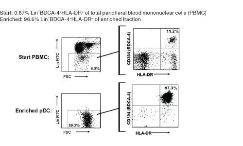
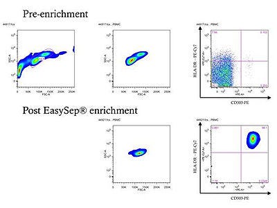
EasySep™ 人浆细胞样树突状细胞富集试剂盒通过免疫磁珠负选,从新鲜的人外周血单个核细胞 (PBMCs) 中分离浆细胞样树突状细胞(pDC)。非目的细胞被识别非pDC的抗体四聚体复合物和葡聚糖包被的磁珠标记。通过EasySep™磁极分离被标记的细胞,无需使用分离柱。目的细胞则被倾倒至一个新的试管中。

磁体兼容性
• EasySep™ Magnet (Catalog #18000) • “The Big Easy” EasySep™ Magnet (Catalog #18001) • EasyPlate™ EasySep™ Magnet (Catalog 18102) • Easy 50 EasySep™ Magnet (Catalog #18002) • RoboSep™-S (Catalog #21000)
 
亚型
细胞分选试剂盒
 
细胞类型
树突状细胞(DCs)
 
种属

 
样本来源
PBMC
 
筛选方法
Negative
 
应用
细胞分选
 
品牌
EasySep,RoboSep
 
研究领域
免疫
 

Data Figures

Typical EasySep™ Human pDC Enrichment Profile

Figure 1. Typical EasySep™ Human pDC Enrichment Profile

Starting with 0.2 - 0.9% pDC in PBMC, the pDC content of the enriched fraction typically ranges from 87 - 97% purity based on the pDC phenotype of Lineage (CD3, CD14, CD16, CD19, CD20, CD34, CD56) negative, HLA-DR positive, and CD304 (BDCA-4) positive.

FACS Purity Data from pDC Enrichment Kit User

Figure 2. FACS Purity Data from pDC Kit User

FACS enrichment plots from Dr. Stuart R. McGregor Dallas of Princeton University. Prior to enrichment, the percentage of pDCs in total PBMC is approximately 0.1% (upper row), however following enrichment, a population of pDCs in excess of 97% pure can be obtained (lower row). pDCs identified using surface marker staining for CD303 and HLA-DR. Data originally posted as part of a product review in Biocompare.

Protocols and Documentation

Find supporting information and directions for use in the Product Information Sheet or explore additional protocols below.

Document Type
Product Name
Catalog #
Lot #
Language
Catalog #
19062
Lot #
All
Language
English
Catalog #
19062RF
Lot #
All
Language
English
Document Type
Safety Data Sheet 1
Catalog #
19062
Lot #
All
Language
English
Document Type
Safety Data Sheet 2
Catalog #
19062
Lot #
All
Language
English
Document Type
Safety Data Sheet 3
Catalog #
19062
Lot #
All
Language
English
Document Type
Safety Data Sheet 4
Catalog #
19062
Lot #
All
Language
English
Document Type
Safety Data Sheet 5
Catalog #
19062
Lot #
All
Language
English
Document Type
Safety Data Sheet 1
Catalog #
19062RF
Lot #
All
Language
English
Document Type
Safety Data Sheet 2
Catalog #
19062RF
Lot #
All
Language
English
Document Type
Safety Data Sheet 3
Catalog #
19062RF
Lot #
All
Language
English
Document Type
Safety Data Sheet 4
Catalog #
19062RF
Lot #
All
Language
English
Document Type
Safety Data Sheet 5
Catalog #
19062RF
Lot #
All
Language
English

Applications

This product is designed for use in the following research area(s) as part of the highlighted workflow stage(s). Explore these workflows to learn more about the other products we offer to support each research area.

Resources and Publications

Educational Materials (8)

Frequently Asked Questions

Can EasySep™ be used for either positive or negative selection?

Yes. The EasySep™ kits use either a negative selection approach by targeting and removing unwanted cells or a positive selection approach targeting desired cells. Depletion kits are also available for the removal of cells with a specific undesired marker (e.g. GlyA).

How does the separation work?

Magnetic particles are crosslinked to cells using Tetrameric Antibody Complexes (TAC). When placed in the EasySep™ Magnet, labeled cells migrate to the wall of the tube. The unlabeled cells are then poured off into a separate fraction.

Which columns do I use?

The EasySep™ procedure is column-free. That's right - no columns!

How can I analyze the purity of my enriched sample?

The Product Information Sheet provided with each EasySep™ kit contains detailed staining information.

Can EasySep™ separations be automated?

Yes. RoboSep™, the fully automated cell separator, automates all EasySep™ labeling and cell separation steps.

Can EasySep™ be used to isolate rare cells?

Yes. We recommend a cell concentration of 2x10⁸ cells/mL and a minimum working volume of 100 µL. Samples containing 2x10⁷ cells or fewer should be suspended in 100 µL of buffer.

Are the EasySep™ magnetic particles FACS-compatible?

Yes, the EasySep™ particles are flow cytometry-compatible, as they are very uniform in size and about 5000X smaller than other commercially available magnetic beads used with column-free systems.

Can the EasySep™ magnetic particles be removed after enrichment?

No, but due to the small size of these particles, they will not interfere with downstream applications.

Can I alter the separation time in the magnet?

Yes; however, this may impact the kit's performance. The provided EasySep™ protocols have already been optimized to balance purity, recovery and time spent on the isolation.

For positive selection, can I perform more than 3 separations to increase purity?

Yes, the purity of targeted cells will increase with additional rounds of separations; however, cell recovery will decrease.

How does the binding of the EasySep™ magnetic particle affect the cells? is the function of positively selected cells altered by the bound particles?

Hundreds of publications have used cells selected with EasySep™ positive selection kits for functional studies. Our in-house experiments also confirm that selected cells are not functionally altered by the EasySep™ magnetic particles.

If particle binding is a key concern, we offer two options for negative selection. The EasySep™ negative selection kits can isolate untouched cells with comparable purities, while RosetteSep™ can isolate untouched cells directly from whole blood without using particles or magnets.

Publications (15)

TLR2 and TLR7 mediate distinct immunopathological and antiviral plasmacytoid dendritic cell responses to SARS-CoV-2 infection. R. M. van der Sluis et al. The EMBO journal 2022 may

Abstract

Understanding the molecular pathways driving the acute antiviral and inflammatory response to SARS-CoV-2 infection is critical for developing treatments for severe COVID-19. Here, we find decreasing number of circulating plasmacytoid dendritic cells (pDCs) in COVID-19 patients early after symptom onset, correlating with disease severity. pDC depletion is transient and coincides with decreased expression of antiviral type I IFN? and of systemic inflammatory cytokines CXCL10 and IL-6. Using an in vitro stem cell-based human pDC model, we further demonstrate that pDCs, while not supporting SARS-CoV-2 replication, directly sense the virus and in response produce multiple antiviral (interferons: IFN? and IFN?1) and inflammatory (IL-6, IL-8, CXCL10) cytokines that protect epithelial cells from de novo SARS-CoV-2 infection. Via targeted deletion of virus-recognition innate immune pathways, we identify TLR7-MyD88 signaling as crucial for production of antiviral interferons (IFNs), whereas Toll-like receptor (TLR)2 is responsible for the inflammatory IL-6 response. We further show that SARS-CoV-2 engages the receptor neuropilin-1 on pDCs to selectively mitigate the antiviral interferon response, but not the IL-6 response, suggesting neuropilin-1 as potential therapeutic target for stimulation of TLR7-mediated antiviral protection.
Human Plasmacytoid Dendritic Cells Express C-Type Lectin Receptors and Attach and Respond to Aspergillus fumigatus. S. D. Maldonado et al. Journal of immunology (Baltimore, Md. : 1950) 2022 aug

Abstract

Plasmacytoid dendritic cells (pDCs) have been implicated as having a role in antifungal immunity, but mechanisms of their interaction with fungi and the resulting cellular responses are not well understood. In this study, we identify the direct and indirect biological response of human pDCs to the fungal pathogen Aspergillus fumigatus and characterize the expression and regulation of antifungal receptors on the pDC surface. Results indicate pDCs do not phagocytose Aspergillus conidia, but instead bind hyphal surfaces and undergo activation and maturation via the upregulation of costimulatory and maturation markers. Measuring the expression of C-type lectin receptors dectin-1, dectin-2, dectin-3, and mannose receptor on human pDCs revealed intermediate expression of each receptor compared with monocytes. The specific dectin-1 agonist curdlan induced pDC activation and maturation in a cell-intrinsic and cell-extrinsic manner. The indirect activation of pDCs by curdlan was much stronger than direct stimulation and was mediated through cytokine production by other PBMCs. Overall, our data indicate pDCs express various C-type lectin receptors, recognize and respond to Aspergillus hyphal Ag, and serve as immune enhancers or modulators in the overarching fungal immune response.
Genomic Multiple Sclerosis Risk Variants Modulate the Expression of the ANKRD55-IL6ST Gene Region in Immature Dendritic Cells. J. Mena et al. Frontiers in immunology 2021

Abstract

Intronic single-nucleotide polymorphisms (SNPs) in the ANKRD55 gene are associated with the risk for multiple sclerosis (MS) and rheumatoid arthritis by genome-wide association studies (GWAS). The risk alleles have been linked to higher expression levels of ANKRD55 and the neighboring IL6ST (gp130) gene in CD4+ T lymphocytes of healthy controls. The biological function of ANKRD55, its role in the immune system, and cellular sources of expression other than lymphocytes remain uncharacterized. Here, we show that monocytes gain capacity to express ANKRD55 during differentiation in immature monocyte-derived dendritic cells (moDCs) in the presence of interleukin (IL)-4/granulocyte-macrophage colony-stimulating factor (GM-CSF). ANKRD55 expression levels are further enhanced by retinoic acid agonist AM580 but downregulated following maturation with interferon (IFN)-$\gamma$ and lipopolysaccharide (LPS). ANKRD55 was detected in the nucleus of moDC in nuclear speckles. We also analyzed the adjacent IL6ST, IL31RA, and SLC38A9 genes. Of note, in healthy controls, MS risk SNP genotype influenced ANKRD55 and IL6ST expression in immature moDC in opposite directions to that in CD4+ T cells. This effect was stronger for a partially correlated SNP, rs13186299, that is located, similar to the main MS risk SNPs, in an ANKRD55 intron. Upon analysis in MS patients, the main GWAS MS risk SNP rs7731626 was associated with ANKRD55 expression levels in CD4+ T cells. MoDC-specific ANKRD55 and IL6ST mRNA levels showed significant differences according to the clinical form of the disease, but, in contrast to healthy controls, were not influenced by genotype. We also measured serum sgp130 levels, which were found to be higher in homozygotes of the protective allele of rs7731626. Our study characterizes ANKRD55 expression in moDC and indicates monocyte-to-dendritic cell (Mo-DC) differentiation as a process potentially influenced by MS risk SNPs.

更多信息

更多信息
Species Human
Magnet Compatibility • EasySep™ Magnet (Catalog #18000) • “The Big Easy” EasySep™ Magnet (Catalog #18001) • EasyPlate™ EasySep™ Magnet (Catalog 18102) • Easy 50 EasySep™ Magnet (Catalog #18002) • RoboSep™-S (Catalog #21000)
Sample Source PBMC
Selection Method Negative
标记抗体
PRODUCTS ARE FOR RESEARCH USE ONLY AND NOT INTENDED FOR HUMAN OR ANIMAL DIAGNOSTIC OR THERAPEUTIC USES UNLESS OTHERWISE STATED. FOR ADDITIONAL INFORMATION ON QUALITY AT STEMCELL, REFER TO WWW.STEMCELL.COM/COMPLIANCE.
版权 © 2025 STEMCELL Technologies 技术有限公司。保留所有权利。