产品号 #19765_C
小鼠Naïve CD4+ T细胞的免疫磁珠负选
小鼠Naïve CD4+ T细胞的免疫磁珠负选
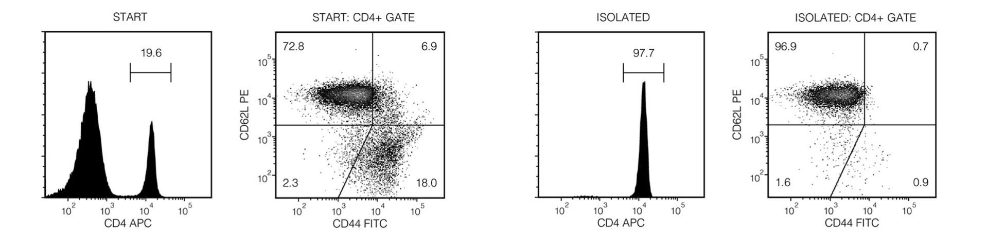
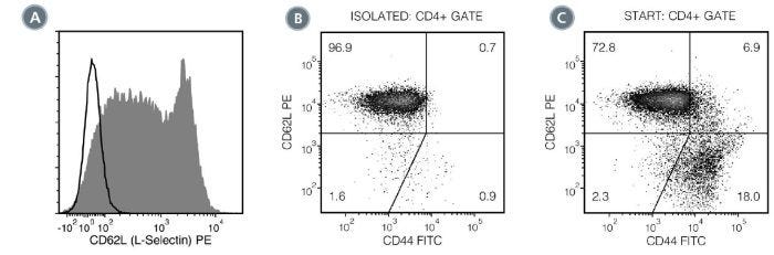
使用EasySep™小鼠初始CD4+ T细胞分选试剂盒,可通过免疫磁珠负选轻松高效地从小鼠脾细胞中分离高纯度初始CD4+ T细胞(CD4+CD44lowCD62Lhigh)。当使用其它类型组织来源的单细胞悬液时,该试剂盒可能需要优化。EasySep™结合了单克隆抗体的特异性和无柱磁性系统的简便性,迄今已广泛应用于发表的研究中超过20年。
在此EasySep™负选过程中,非目的细胞通过抗体复合物与磁珠标记。通过EasySep™磁极将被磁珠标记的细胞与未被标记的目的初始CD4+ T细胞(CD4+CD44lowCD62Lhigh)细胞分离,接着简单地将目的细胞倾倒或吸取至一个新的试管中即可。仅需15分钟磁珠分选后, 获得的初始CD4+ T细胞可直接用于流式细胞术、细胞培养或基于细胞的实验 等下游应用。
了解更多关于免疫磁珠EasySep™技术的工作原理,或如何通过RoboSep™实现全自动化免疫磁珠细胞分选 。探索更多优化您实验流程的产品,包括培养基、添加剂、抗体等。
磁体兼容性
• EasySep™ Magnet (Catalog #18000)
• “The Big Easy” EasySep™ Magnet (Catalog #18001)
• EasyEights™ EasySep™ Magnet (Catalog #18103)
• RoboSep™-S (Catalog #21000)
亚型
细胞分选试剂盒
细胞类型
T 细胞,T 细胞,CD4+
种属
小鼠
样本来源
其它细胞系,Spleen
筛选方法
Negative
应用
细胞分选
品牌
EasySep,RoboSep
研究领域
免疫
Find supporting information and directions for use in the Product Information Sheet or explore additional protocols below.
This product is designed for use in the following research area(s) as part of the highlighted workflow stage(s). Explore these workflows to learn more about the other products we offer to support each research area.
| Species | Mouse |
|---|---|
| Magnet Compatibility | • EasySep™ Magnet (Catalog #18000) • “The Big Easy” EasySep™ Magnet (Catalog #18001) • EasyEights™ EasySep™ Magnet (Catalog #18103) • RoboSep™-S (Catalog #21000) |
| Sample Source | Other, Spleen |
| Selection Method | Negative |
小鼠T细胞激活和扩增试剂
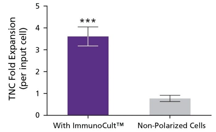
利用免疫磁珠负选技术,27.5分钟完成细胞分选
不带标记的小鼠T细胞免疫磁珠负选
免疫磁珠负选不带标记的小鼠CD4+ T细胞
抗小鼠CD62L(L-选择素)的大鼠单克隆IgG2a抗体
大鼠单克隆IgG2a抗体,抗小鼠CD4