产品号 #19851_C
不带标记的小鼠T细胞免疫磁珠分选
不带标记的小鼠T细胞免疫磁珠分选
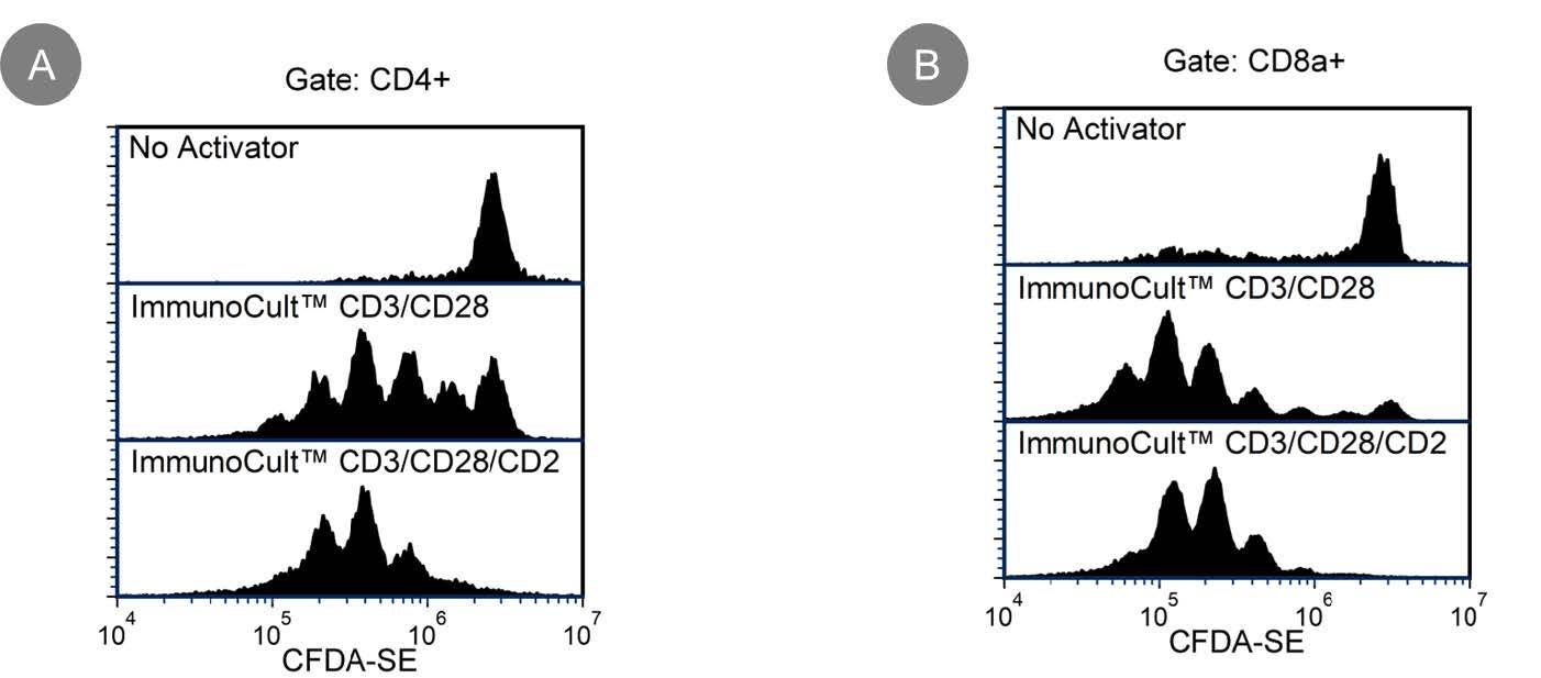
使用EasySep™小鼠T细胞分选试剂盒,通过免疫磁珠负选,可简便高效地从脾细胞或其他组织的单细胞悬液中分离高纯度的小鼠T细胞。EasySep™技术结合单克隆抗体的特异性和无柱磁分选系统的简便性,已在发表的研究中广泛应用超过20年。
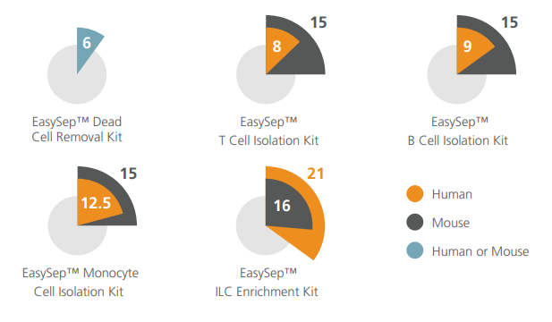
在该EasySep™负选流程中,非目的细胞会被抗体复合物与磁珠标记。表达以下标志物的非目的细胞将被识别并去除:CD11b、CD45R、Ter119、CD49b、CD19和CD24。使用EasySep™磁极吸附后,通过简单地将目的细胞倾倒或吸取至一个新的试管中,即可将被磁珠标记的细胞与不带标记的目的细胞分离开来。在短至15分钟的磁珠分选后,目的T细胞可立即用于流式细胞术、培养及基于细胞的实验等下游应用。
了解更多EasySep™免疫磁珠技术的工作原理,或如何通过RoboSep™实现全自动化免疫磁珠细胞分选。探索为您的实验流程优化的更多产品,包括培养基、补充剂、抗体等。
磁体兼容性
• EasySep™ Magnet (Catalog #18000)
• “The Big Easy” EasySep™ Magnet (Catalog #18001)
• EasyEights™ EasySep™ Magnet (Catalog #18103)
• RoboSep™-S (Catalog #21000)
亚型
细胞分选试剂盒
细胞类型
T 细胞
种属
小鼠
样本来源
其它细胞系,Spleen
筛选方法
Negative
应用
细胞分选
品牌
EasySep,RoboSep
研究领域
免疫
Find supporting information and directions for use in the Product Information Sheet or explore additional protocols below.
This product is designed for use in the following research area(s) as part of the highlighted workflow stage(s). Explore these workflows to learn more about the other products we offer to support each research area.
| Species | Mouse |
|---|---|
| Magnet Compatibility | • EasySep™ Magnet (Catalog #18000) • “The Big Easy” EasySep™ Magnet (Catalog #18001) • EasyEights™ EasySep™ Magnet (Catalog #18103) • RoboSep™-S (Catalog #21000) |
| Sample Source | Other, Spleen |
| Selection Method | Negative |
15分钟免疫磁珠负选试剂盒
免疫磁珠负选不带标记的小鼠CD4+ T细胞
通过免疫磁珠负选分离无磁珠标记的小鼠CD8+ T细胞
细胞分选缓冲液
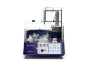
全自动细胞分选仪器
亚美尼亚仓鼠单克隆IgG1抗体,抗小鼠CD3e