EasySep™ Direct人总淋巴细胞分选试剂盒

直接从全血中免疫磁珠负选人总淋巴细胞

产品号 #(选择产品)

产品号 #19655_C

直接从全血中免疫磁珠负选人总淋巴细胞

产品优势

  • > 99.9%的红细胞去除率,无需密度梯度离心、沉降或裂解
  • 操作简单、快捷,且无需分离柱
  • 分选获得的细胞纯度高达96%
  • 分选得到的细胞不带标记

产品组分包括

  • EasySep™ Direct人总淋巴细胞分选试剂盒(产品号 #19655)
    • EasySep™ Direct人总淋巴细胞分选抗体混合物, 2 x 2.5 mL
    • EasySep™ Direct RapidSpheres™ 磁珠, 4 x 2.5 mL
  • EasySep™ Direct人总淋巴细胞分选试剂盒(用于RoboSep™)(产品号 #19655RF)
    • EasySep™ Direct人总淋巴细胞分选抗体混合物, 2 x 2.5 mL
    • EasySep™ Direct RapidSpheres™ 磁珠, 4 x 2.5 mL
    • RoboSep™ 缓冲液(产品号 #20104)
    • RoboSep™过滤吸头(产品号 #20125)
New look, same high quality and support! You may notice that your instrument or reagent packaging looks slightly different from images displayed on the website, or from previous orders. We are updating our look but rest assured, the products themselves and how you should use them have not changed. Learn more

总览

使用EasySep™ Direct 人总淋巴细胞分选试剂盒,可轻松高效地从人全血、白膜层或脾脏样本中通过免疫磁珠负选获得高纯度的人总淋巴细胞。EasySep™技术结合单克隆抗体的特异性和免磁柱系统的简便性,已在发表的研究中广泛应用超过20年。

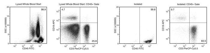
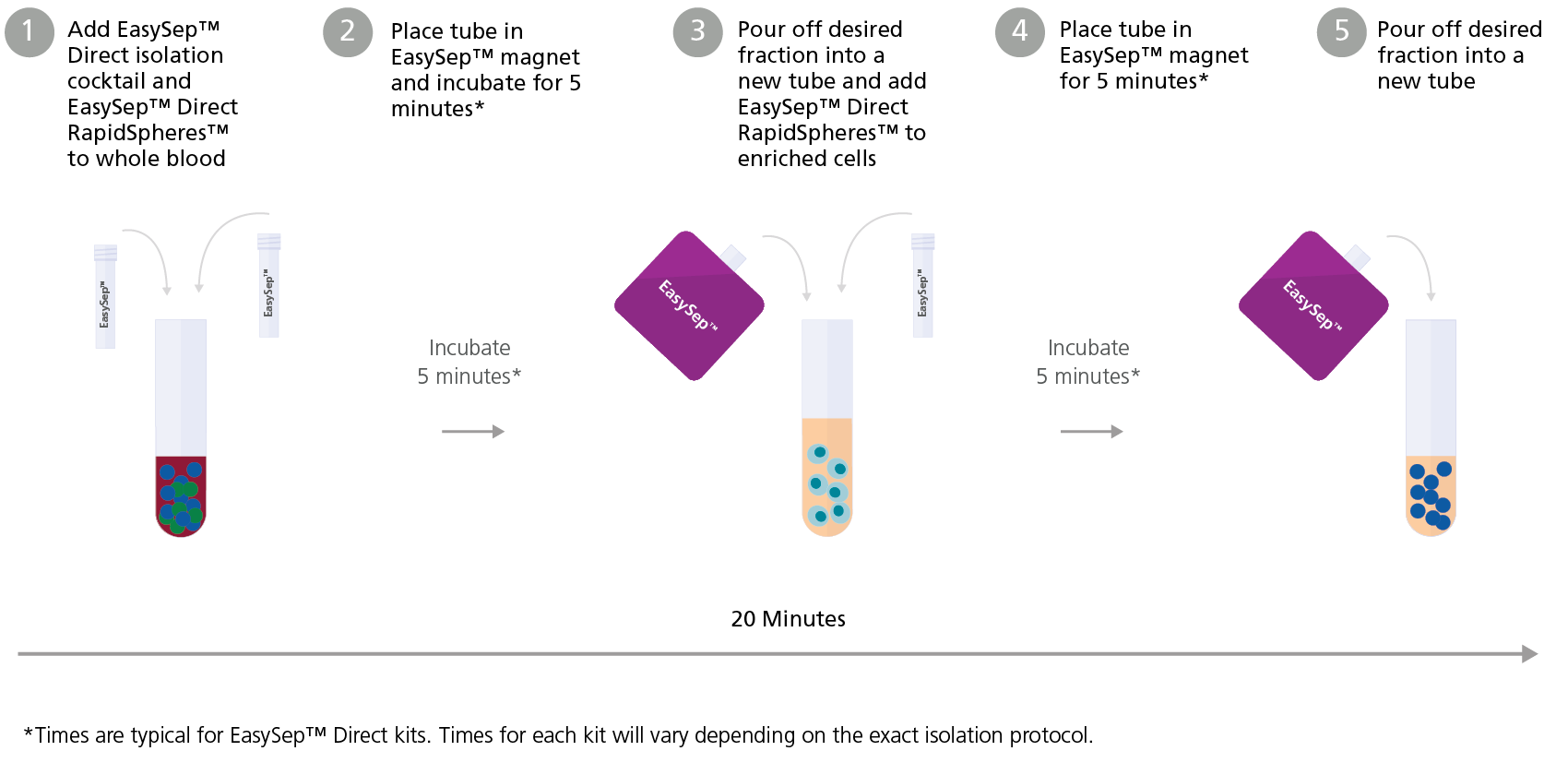
在该EasySep™负选流程中,非目的细胞会被抗体复合物和EasySep™ Direct RapidSpheres™磁珠标记。以下非目的细胞会被非特异性去除:单核细胞、中性粒细胞、嗜酸性粒细胞和血小板。通过EasySep™磁极将被磁珠标记的细胞与未被标记的目的细胞分离,目的细胞可通过倾倒或移液收集到新试管中,分选后的细胞可立即用于流式细胞术、细胞培养或DNA/RNA提取等下游应用。

了解更多EasySep™免疫磁珠技术的工作原理,或者如何通过RoboSep™实现全自动化免疫磁珠细胞分选。探索更多为您的实验流程优化的产品,包括细胞鉴定、冷冻保存等相关试剂。

磁体兼容性
• EasySep™ Magnet (Catalog #18000) • “The Big Easy” EasySep™ Magnet (Catalog #18001) • Easy 50 EasySep™ Magnet (Catalog #18002) • EasyEights™ EasySep™ Magnet (Catalog #18103) • RoboSep™-S (Catalog #21000)
 
亚型
细胞分选试剂盒
 
细胞类型
淋巴细胞
 
种属

 
样本来源
Whole Blood
 
筛选方法
Negative
 
应用
细胞分选
 
品牌
EasySep
 
研究领域
嵌合体,HLA,免疫
 

Data Figures

EasySep™ Direct Human Total Lymphocyte Isolation Profile

Figure 1. Typical EasySep™ Direct Human Total Lymphocyte Isolation Profile

Starting with human whole blood from normal healthy donors, the typical total lymphocyte (CD3-CD19+ and CD3+) content of the non-lysed final isolated fraction is 96.7 ± 1.5% (gated on CD45) or 95.8 ± 2.2% (not gated on CD45). In the example above, the total lymphocyte (CD3-CD19+ and CD3+) content of the lysed whole blood start sample and non-lysed final isolated fraction is 30.5% and 93.2% (gated on CD45), respectively, or 30.2% and 93.0% (not gated on CD45), respectively. The starting frequency of total lymphocytes in the non-lysed whole blood start sample above is 0.055% (data not shown).

Using EasySep™ Direct Increases Lymphocyte Purity While Maintaining Cell Yield

Figure 2. Using EasySep™ Direct Increases Lymphocyte Purity While Maintaining Cell Yield

Cells were isolated from the same deceased donor whole blood sample using (A) a density gradient separation medium or (B) EasySep™ Direct Human Total Lymphocyte Isolation Kit (Catalog #19655) and analyzed by flow cytometry. Cleaner samples were obtained with EasySep™ Direct. (C) Samples isolated using EasySep™ Direct contained fewer contaminating cells, yet maintained overall numbers of T (CD3+) and B (CD19+) cells comparable to samples isolated using the density gradient medium. Each column with error bar represents the mean ± SD from 24 mL whole blood (n = 20 donors). Data kindly provided by Dr. Robert Liwski and originally presented in the technical bulletin on Cell Separation Solutions for the Flow Cytometric Crossmatch Assay.

Use of Highly Enriched Lymphocytes Isolated with EasySep™ Direct Improves DSA Detection Compared to Whole Leukocyte Cell Preparations

Figure 3. Use of Highly Enriched Lymphocytes Isolated with EasySep™ Direct Improves DSA Detection Compared to Whole Leukocyte Cell Preparations

Lymphocytes (Ly), neutrophils (Nu), and monocytes (Mo) were isolated from volunteer donors (n = 5) using EasySep™ Direct Catalog #19655, #19666, and #19669, respectively. Whole leukocyte (WL) preparations were obtained by adding Ly, Nu, and Mo cells in equal proportions. WL (low lymphocyte purity) and Ly (high lymphocyte purity) preparations were treated with pronase and then used to perform the flow cytometry cross-match (FCXM) assay against negative control sera or several dilutions of positive control sera. The median channel fluorescence shifts (MCFS) were generated by using the negative control sera samples as a baseline. The MCF shifts between WL and Ly were then compared. Each column with error bars represents the mean ± SEM (n = 5 donors). Data kindly provided by Dr. Robert Liwski and originally presented in the technical bulletin on Cell Separation Solutions for the Flow Cytometric Crossmatch Assay.

Typical EasySep™ Direct Protocol

Figure 4. Typical EasySep™ Direct Protocol

Protocols and Documentation

Find supporting information and directions for use in the Product Information Sheet or explore additional protocols below.

Document Type
Product Name
Catalog #
Lot #
Language
Catalog #
19655
Lot #
All
Language
English
Catalog #
19655RF
Lot #
All
Language
English
Document Type
Safety Data Sheet 1
Catalog #
19655
Lot #
All
Language
English
Document Type
Safety Data Sheet 2
Catalog #
19655
Lot #
All
Language
English
Document Type
Safety Data Sheet 1
Catalog #
19655RF
Lot #
All
Language
English
Document Type
Safety Data Sheet 2
Catalog #
19655RF
Lot #
All
Language
English
Document Type
Safety Data Sheet 3
Catalog #
19655RF
Lot #
All
Language
English

Applications

This product is designed for use in the following research area(s) as part of the highlighted workflow stage(s). Explore these workflows to learn more about the other products we offer to support each research area.

Resources and Publications

Educational Materials (19)

Frequently Asked Questions

Can EasySep™ be used for either positive or negative selection?

Yes. The EasySep™ kits use either a negative selection approach by targeting and removing unwanted cells or a positive selection approach targeting desired cells. Depletion kits are also available for the removal of cells with a specific undesired marker (e.g. GlyA).

How does the separation work?

Magnetic particles are crosslinked to cells using Tetrameric Antibody Complexes (TAC). When placed in the EasySep™ Magnet, labeled cells migrate to the wall of the tube. The unlabeled cells are then poured off into a separate fraction.

Which columns do I use?

The EasySep™ procedure is column-free. That's right - no columns!

How can I analyze the purity of my enriched sample?

The Product Information Sheet provided with each EasySep™ kit contains detailed staining information.

Can EasySep™ separations be automated?

Yes. RoboSep™, the fully automated cell separator, automates all EasySep™ labeling and cell separation steps.

Can EasySep™ be used to isolate rare cells?

Yes. We recommend a cell concentration of 2x10⁸ cells/mL and a minimum working volume of 100 µL. Samples containing 2x10⁷ cells or fewer should be suspended in 100 µL of buffer.

Are the EasySep™ magnetic particles FACS-compatible?

Yes, the EasySep™ particles are flow cytometry-compatible, as they are very uniform in size and about 5000X smaller than other commercially available magnetic beads used with column-free systems.

Can the EasySep™ magnetic particles be removed after enrichment?

No, but due to the small size of these particles, they will not interfere with downstream applications.

Can I alter the separation time in the magnet?

Yes; however, this may impact the kit's performance. The provided EasySep™ protocols have already been optimized to balance purity, recovery and time spent on the isolation.

For positive selection, can I perform more than 3 separations to increase purity?

Yes, the purity of targeted cells will increase with additional rounds of separations; however, cell recovery will decrease.

How does the binding of the EasySep™ magnetic particle affect the cells? is the function of positively selected cells altered by the bound particles?

Hundreds of publications have used cells selected with EasySep™ positive selection kits for functional studies. Our in-house experiments also confirm that selected cells are not functionally altered by the EasySep™ magnetic particles.

If particle binding is a key concern, we offer two options for negative selection. The EasySep™ negative selection kits can isolate untouched cells with comparable purities, while RosetteSep™ can isolate untouched cells directly from whole blood without using particles or magnets.

Publications (5)

Ageing-resembling phenotype of long-term allogeneic hematopoietic cells recipients compared to their donors. M. C. Czarnog\'orski et al. Immunity & ageing : I & A 2022 nov

Abstract

BACKGROUND Ageing is a complex phenomenon that leads to decreased proliferative activity, loss of function of the cells, and cellular senescence. Senescence of the immune system exacerbates individual's immune response, both humoral and cellular but increases the frequency of infections. We hypothesized that physiological ageing of adaptive immune system occurs in recipients of allogeneic hematopoietic cells transplant (allo-HCT) at faster rate when compared to their respective donors since the small number of donor cells undergo immense proliferative stress restoring recipients hematopoiesis. We compared molecular characterizations of ageing between recipients and donors of allo-HCT: telomeric length and immunophenotypic changes in main lymphocyte subsets - CD4+, CD8+, CD19+, CD56+. RESULTS Median telomeric length (TL) of CD8+ lymphocytes was significantly longer in donors compared to recipients (on average 2,1 kb and 1,7 kb respectively, p??=??0,02). Similar trends were observed for CD4+ and CD19+ although the results did not reach statistical significance. We have also found trends in the immunophenotype between recipients and donors in the subpopulations of CD4+ (na{\{i}}ve and effector memory) CD8+ Eomes+ and B-lymphocytes (B1 and B2). Lower infection risk recipients had also a significantly greater percentage of NK cells (22 3%) than high-risk patients (9 3%) p??=??0 04. CONCLUSION Our data do not support the initial hypothesis of accelerated aging in the long term all-HCT recipients with the exception of the recipients lymphocytes (mainly CD8+) which present some molecular features characteristic for physiological ageing (telomeric shortening immunophenotype) when compared to their respective donors. However a history of lower infection numbers in HCT recipients seems to be associated with increased percentage of NK cells. The history of GVHD seems not to affect the rate of ageing. Therefore it is safe to conclude that the observed subtle differences between recipients' and donors' cells result mainly from the proliferative stress in the early period after allo-HCT and the difference between hosts' and recipients' microenvironments."
Dexamethasone modulates immature neutrophils and interferon programming in severe COVID-19. S. Sinha et al. Nature medicine 2022 jan

Abstract

Although critical for host defense, innate immune cells are also pathologic drivers of acute respiratory distress syndrome (ARDS). Innate immune dynamics during Coronavirus Disease 2019 (COVID-19) ARDS, compared to ARDS from other respiratory pathogens, is unclear. Moreover, mechanisms underlying the beneficial effects of dexamethasone during severe COVID-19 remain elusive. Using single-cell RNA sequencing and plasma proteomics, we discovered that, compared to bacterial ARDS, COVID-19 was associated with expansion of distinct neutrophil states characterized by interferon (IFN) and prostaglandin signaling. Dexamethasone during severe COVID-19 affected circulating neutrophils, altered IFNactive neutrophils, downregulated interferon-stimulated genes and activated IL-1R2+ neutrophils. Dexamethasone also expanded immunosuppressive immature neutrophils and remodeled cellular interactions by changing neutrophils from information receivers into information providers. Male patients had higher proportions of IFNactive neutrophils and preferential steroid-induced immature neutrophil expansion, potentially affecting outcomes. Our single-cell atlas (see 'Data availability' section) defines COVID-19-enriched neutrophil states and molecular mechanisms of dexamethasone action to develop targeted immunotherapies for severe COVID-19.
Cutting through the weeds: Evaluation of a novel adsorption with crossmatch cells and elution protocol to sharpen HLA antibody identification by the single antigen bead assay. R. S. Liwski et al. Frontiers in genetics 2022

Abstract

The single antigen bead (SAB) assay is the most used test for the identification of HLA specific antibodies pre- and post-transplant. Nevertheless, detection of spurious reactivities remains a recognized assay limitation. In addition, the presence of weak reactivity patterns can complicate unacceptable antigen assignment. This work presents the evaluation of the adsorption with crossmatch cells and elution (AXE) technique, which was designed to help differentiate weak HLA specific antibodies targeting native antigens from spurious and background SAB assay reactivity. The AXE protocol uses selected donor cells to adsorb HLA specific antibodies from sera of interest. Bound antibodies are then eluted off washed cells and identified using the SAB assay. Only antibodies targeting native HLA are adsorbed. Assay evaluation was performed using five cell donors and pooled positive control serum. AXE efficiency was determined by comparing SAB reactivity of adsorbed/eluted antibody to that of the antibodies in unadsorbed sera. A robust efficiency was seen across a wide range of original MFI for donor specific antibodies (DSA). A higher absorption/elution recovery was observed for HLA class I antigens vs. class II. Locus-specific variation was also observed, with high-expression HLA loci (HLA-A/B/DR) providing the best recovery. Importantly, negligible reactivity was detected in the last wash control, confirming that AXE eluates were not contaminated with HLA antibody carry-over. Donor cells incubated with autologous and DSA-containing allogeneic sera showed that AXE selectively adsorbed HLA antibodies in a donor antigen-specific manner. Importantly, antibodies targeting denatured epitopes or other non-HLA antigens were not detected by AXE. AXE was particularly effective at distinguishing weak HLA antibodies from background reactivity. When combined with epitope analysis, AXE enhanced precise identification of antibody-targeted eplets and even facilitated the characterization of a potential novel eplet. Comparison of AXE to flow cytometric crossmatching further revealed that AXE was a more sensitive technique in the detection of weak DSA. Spurious reactivities on the current SAB assay have a deleterious impact on the assignment of clinically relevant HLA specificities. The AXE protocol is a novel test that enables users to interrogate reactive patterns of interest and discriminate HLA specific antibodies from spurious reactivity.

更多信息

更多信息
Species Human
Magnet Compatibility • EasySep™ Magnet (Catalog #18000) • “The Big Easy” EasySep™ Magnet (Catalog #18001) • Easy 50 EasySep™ Magnet (Catalog #18002) • EasyEights™ EasySep™ Magnet (Catalog #18103) • RoboSep™-S (Catalog #21000)
Sample Source Whole Blood
Selection Method Negative
标记抗体
PRODUCTS ARE FOR RESEARCH USE ONLY AND NOT INTENDED FOR HUMAN OR ANIMAL DIAGNOSTIC OR THERAPEUTIC USES UNLESS OTHERWISE STATED. FOR ADDITIONAL INFORMATION ON QUALITY AT STEMCELL, REFER TO WWW.STEMCELL.COM/COMPLIANCE.
版权 © 2025 STEMCELL Technologies 技术有限公司。保留所有权利。