Figure 1. Human iPSC-Derived Retinal Pigment Epithelial Cells Show Characteristic RPE Morphology
Cryopreserved Human iPSC-Derived Retinal Pigment Epithelial (RPE) Cells were thawed and plated onto Corning® Matrigel®-coated plates at 150,000 cells/cm². RPE cells were maintained in STEMdiff™-XF RPE Maturation Medium supplemented with STEMdiff™-ACF RPE Plating Supplement for 7 days, followed by STEMdiff™-XF RPE Maturation Medium alone thereafter. The cells were incubated at 37°C and subsequently analyzed by brightfield microscopy at various time points. Representative microscopy images show that RPE cells display the expected morphology after more than 63 days of culture.
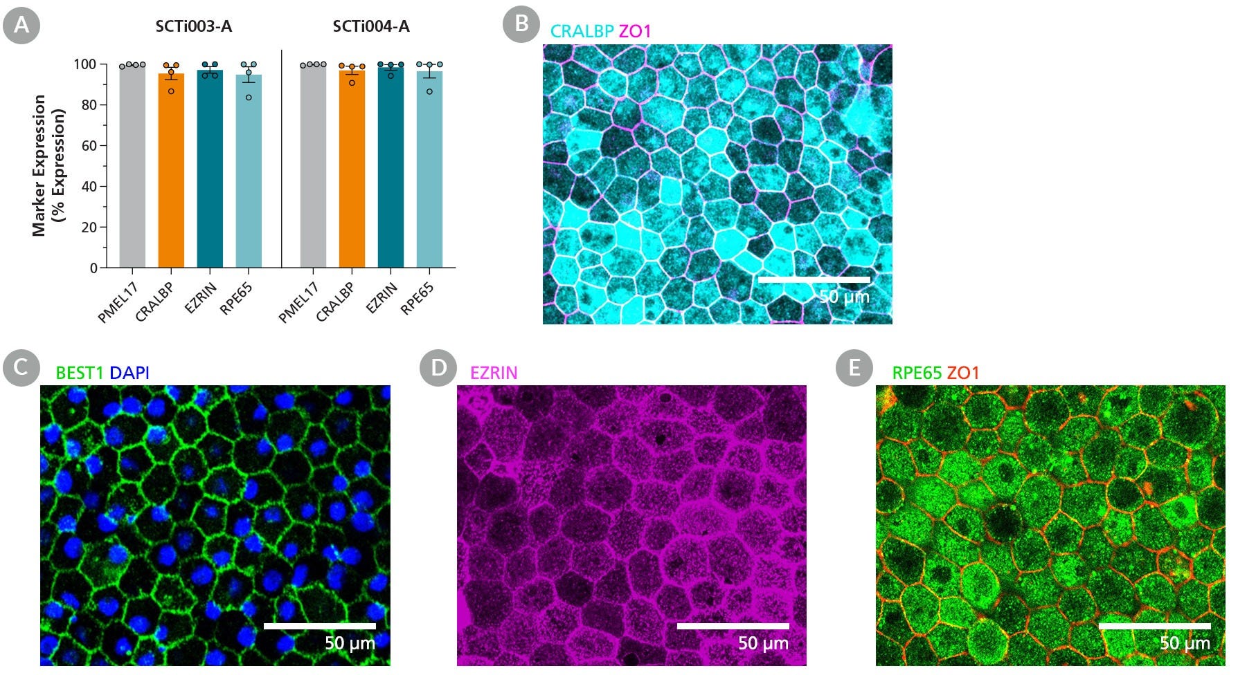
Figure 2. Human iPSC-Derived Retinal Pigment Epithelial (RPE) Cells Are Mature and Functional at Day 35.
Human iPSC-Derived RPE Cells were thawed and cultured in STEMdiff™-XF RPE Maturation Medium for at least 35 days to demonstrate RPE maturity.
(A) The percentage of RPE cells, derived from two different iPSC lines, expressing PMEL17, CRALBP, EZRIN, and RPE65 was assessed by flow cytometry analysis. Data are reported as mean ± SEM (n = 4).
(B,C) Mature RPE cells exhibit high expression of CRALBP and display extensive tight junctions, as indicated by the localization of ZO-1 and BEST1 at cell boundaries. (D,E) Mature RPE cells are polarized, expressing EZRIN (3D projection) and proteins essential for the visual cycle, such as RPE65. These markers are visualized by fluorescence microscopy.
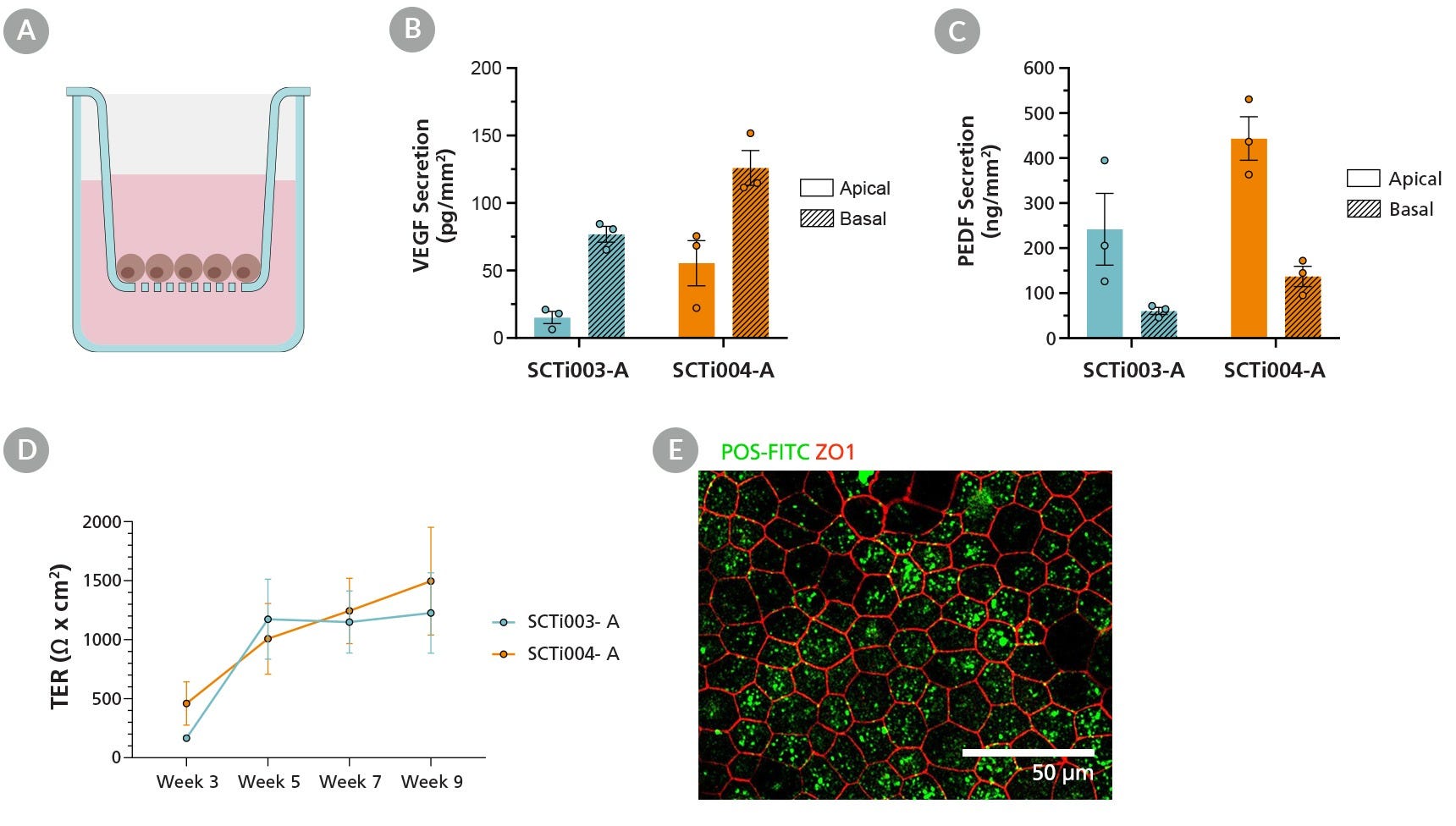
Human iPSC-Derived Retinal Pigment Epithelial (RPE) Cells were cultured on cell culture inserts in STEMdiff™-XF RPE Maturation Medium for 35 days. Apical and basal conditioned media were collected from mature RPE cells, and a sandwich ELISA was performed to quantify vascular endothelial growth factor (VEGF) and pigment epithelium-derived growth factor (PEDF) secretion.
(A) A cross-sectional schematic of the cell insert culture system illustrates the setup. Mature RPE cells, derived from two iPSC lines, secreted higher levels of (B) VEGF basally and (C) PEDF apically, demonstrating correct apicobasal polarity.
(D) Mature RPE cells also generated a strong barrier with high transepithelial resistance (TER). (E) Additionally, mature RPE cells were fed FITC-labeled bovine photoreceptor outer segments (POS) for 4 - 5 hours and efficiently internalized the bovine POS. Data are presented as mean ± SEM.
Thank you for your interest in IntestiCult™ Organoid Growth Medium (Human). Please provide us with your contact information and your local representative will contact you with a customized quote. Where appropriate, they
can also assist you with a(n):
Estimated delivery time for your area
Product sample or exclusive offer
In-lab demonstration
By submitting this form, you are providing your consent to STEMCELL Technologies Canada Inc. and its subsidiaries and affiliates (“STEMCELL”) to collect and use your information, and send you newsletters and emails in accordance with our
privacy policy. Please contact us with any questions that you may have. You can unsubscribe or change your email preferences at any time.
Legal Statement: These iPSCs and their modifications (including but not limited to derivatives or differentiated progeny) shall not be used or administered in (1) human subjects for human clinical use; (2) animals for veterinary use for therapeutic, diagnostic, or prophylactic purposes or (3) any subject in relation to clinical applications, cell therapy, transplantation, and/or regenerative medicines, without limiting the generality of the foregoing. These iPSCs and their modifications (including but not limited to derivatives or differentiated progeny) may not be used for monetization or commercialization purposes, including without limitation, used to, or with the goal to, perform services or supply products or rights, including in the manufacture of cellular therapies or other therapeutics, for monetary gain or the generation of royalties, revenues, sales or other valuable consideration. For clarity, these iPSCs and their modifications (including but not limited to derivatives or differentiated progeny) may not be used for screening compounds, antibodies, proteins or peptides, except for the purposes of target discovery, target validation, or assay development, provided such activities and the results of such activities are not further used for monetization or commercialization purposes. It may be possible to obtain a further license for the prohibited uses referred to in this Limited Use License. Please contact iPSCrequests@stemcell.com for more details. PRODUCTS ARE FOR RESEARCH USE ONLY AND NOT INTENDED FOR HUMAN OR ANIMAL DIAGNOSTIC OR THERAPEUTIC USES UNLESS OTHERWISE STATED.