产品号 #100-1367_C
从人多能干细胞生成未成熟视网膜色素上皮的动物无组分分化试剂盒
Animal component-free supplement for improving survival and attachment of hPSC-derived retinal pigment epithelial cells
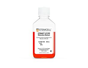
Serum-free and xeno-free medium for generating fully functional and mature hPSC-derived retinal pigment epithelium
Compatible antibodies for purity assessment of isolated cells
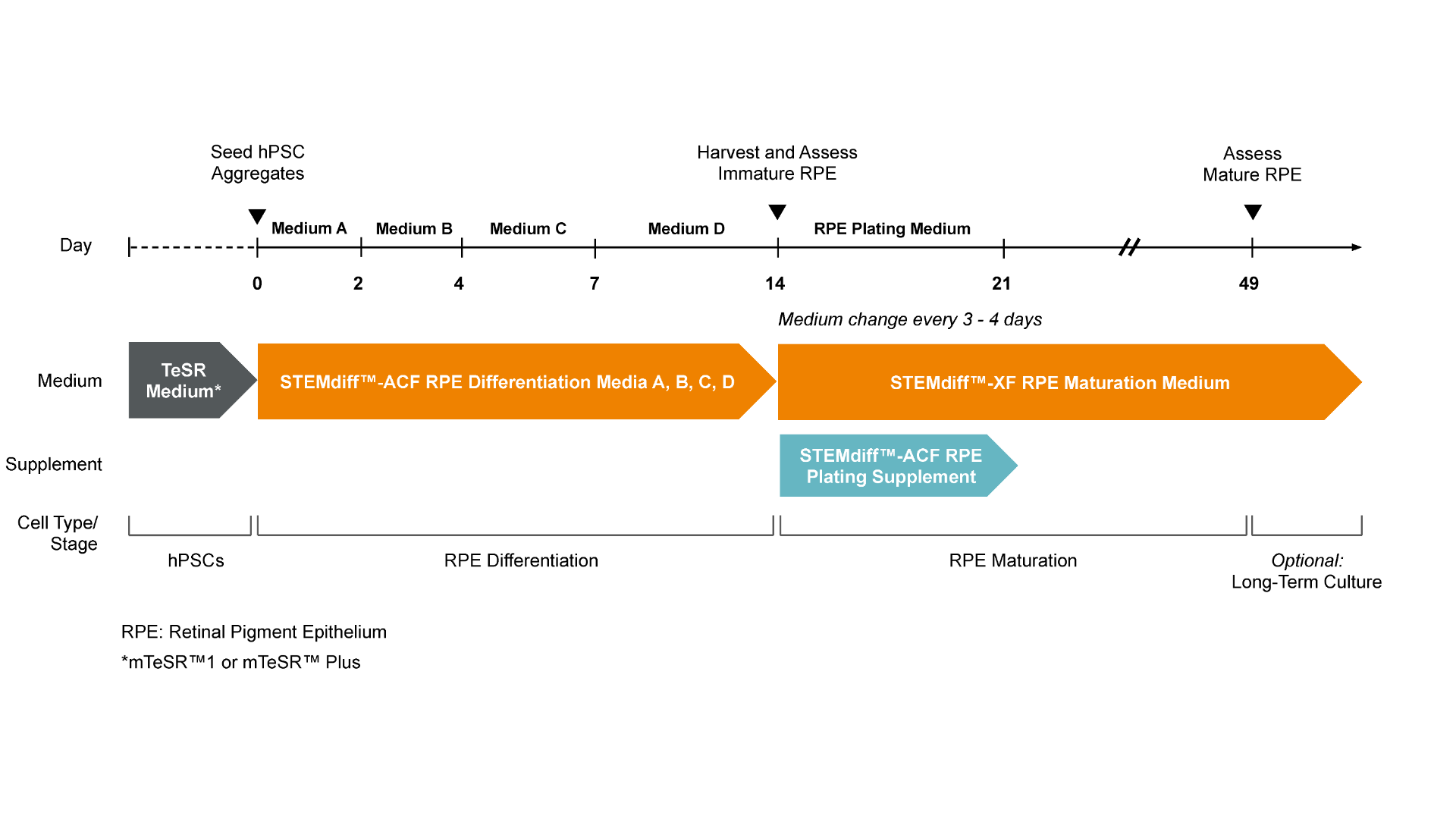
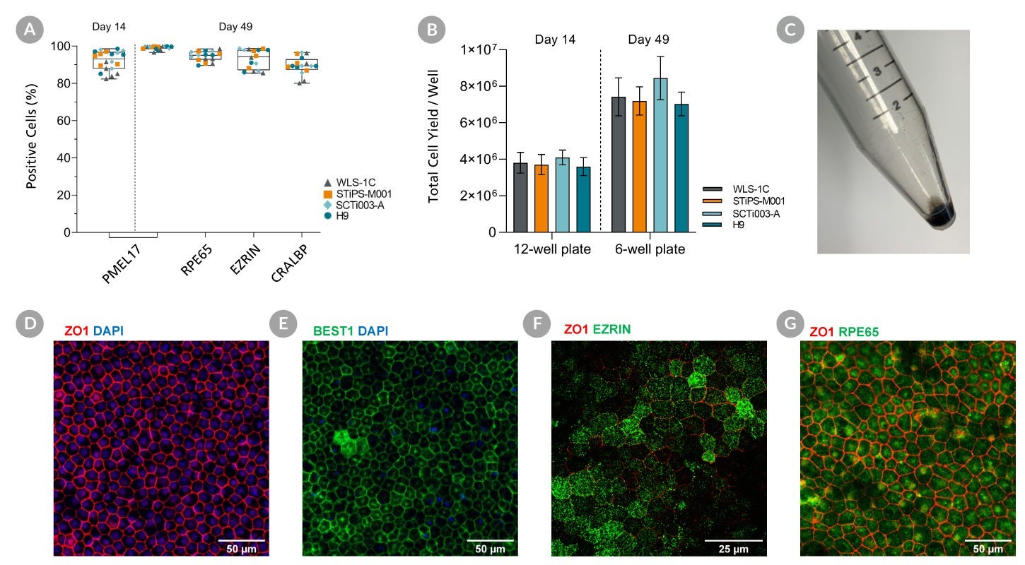
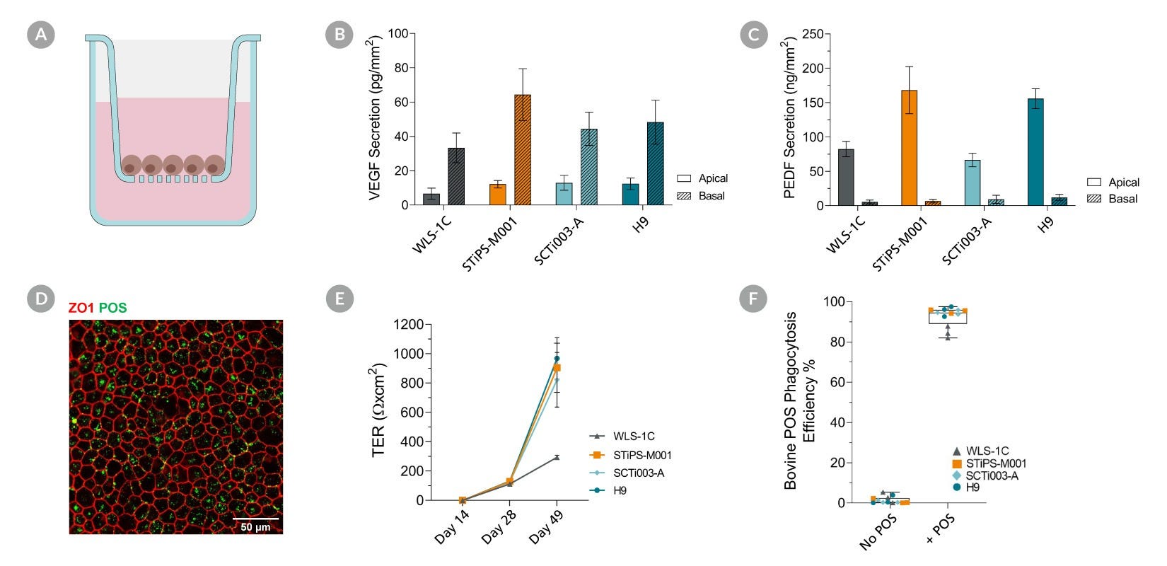
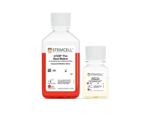
STEMdiff™-ACF RPE分化试剂盒在14天内快速、稳定地生成hpsc来源的未成熟视网膜色素上皮(RPE)。这种不含动物成分和血清的试剂盒从维持在细胞中的成团培养的hPSCs中产生未成熟的RPE (bbb50 % PMEL17+)mTeSR™1或mTeSR™+.
使用STEMdiff™-ACF RPE分化试剂盒生成的未成熟RPE可作为中间细胞库冷冻保存,或使用STEMdiff™-ACF RPE分化试剂盒进一步成熟为功能性RPESTEMdiff™-XF RPE成熟培养基。通过将这些产品一起使用,可以在49天内从人造血干细胞中产生高纯度的RPE群体(图2),而无需手动选择或细胞富集。增加你的RPE细胞分化和成熟的产量和存活率STEMdiff™-ACF RPE电镀补充物这个工作流。
使用这些产品衍生的细胞可用于模拟人类视网膜发育和疾病,药物筛选,细胞和基因治疗验证以及高级组织模型开发。
如果您打算将本产品用于商业或临床应用,请与我们联系。
Cell Type
Neural Cells, PSC-Derived, Pluripotent Stem Cells
Application
Cell Culture, Differentiation
Brand
STEMdiff
Area of Interest
Disease Modeling, Drug Discovery and Toxicity Testing, Neuroscience, Cell Therapy Development
Figure 1. Workflow for the Differentiation of hPSCs into Retinal Pigment Epithelium (RPE) with STEMdiff™ RPE Culture System
hPSC colonies, previously harvested and seeded as clumps, are put directly into the medium provided in STEMdiff™-ACF RPE Differentiation Kit. Seeding into STEMdiff™ RPE Differentiation Medium A induces cells toward immature RPE. A full medium change is performed on Day 1 with fresh STEMdiff™ RPE Differentiation Medium A and then on Day 2 with STEMdiff™ RPE Differentiation Medium B. On Days 4 and 6, medium changes are performed with fresh STEMdiff™ RPE Differentiation Medium C. On Days 7 and every second day thereafter a medium change is performed with fresh STEMdiff™ RPE Differentiation Medium D. On Day 14, immature RPE cells are enzymatically harvested and subcultured in STEMdiff™-XF RPE Maturation Medium with the addition of STEMdiff™-ACF RPE Plating Supplement from Days 14 to 21 to improve survival after passaging. RPE cells begin to mature over the course of 5 weeks of culture in STEMdiff™-XF RPE Maturation Medium and become fully matured by Day 49, possessing key characteristics such as polygonal shape, polarization, pigmentation and phagocytosis.
Figure 2. Robust and Rapid Generation of Mature Retinal Pigment Epithelial Cells (RPE) across multiple PSC Cell Lines with the STEMdiff™-ACF RPE Differentiation Kit
hPSCs were cultured for 14 Days using STEMdiff™-ACF RPE Differentiation Kit and subsequently subcultured in STEMdiff™-XF RPE Maturation Medium. Flow cytometry expression of RPE markers are shown at Day 14 and Day 49. (A) The percentage of cells expressing PMEL17, RPE65, EZRIN, and CRALBP and (B) Viable cell yields for 4 hPSC cell lines. PMEL17 is expressed at Day 14 and 49 while the other markers are only present at Day 49. Data are reported as mean + SEM; n = 16 -20. (C) A cell pellet of mature RPE cells demonstrates the pigmentation. Maturation is further demonstrated with immunohistochemistry for expression of RPE markers at Day 49. (D, E, F, G) Mature RPE display tight junctions marked by localization of ZO1 and BEST1 to cell junctions. Mature RPE are polarized, expressing EZRIN apically and ZO1 subapically and express proteins required for the visual cycle (RPE65).
Figure 3. Mature Retinal Pigment Epithelial (RPE) Cells Display Key Functionalities Corresponding to RPE Behaviour
hPSC’s were cultured for 14 Days using STEMdiff™-ACF RPE Differentiation Kit and subsequently subcultured on cell culture inserts in STEMdiff™-XF RPE Maturation Medium for 5 weeks. Apical and basal conditioned medium were collected from Mature RPE, and a sandwich ELISA was performed to quantify Vascular Endothelial Growth Factor (VEGF) and Pigment Epithelial Derived Growth Factor (PEDF) secretion. (A, B) Mature RPE secreted more basal VEGF and apical PEDF demonstrating RPE display correct apicobasal polarity. Data shown as mean + SEM; n = 3. (C) Mature RPE were able to generate a strong barrier with high transepithelial resistance (TER). Data shown as mean + SEM; n = 3-6. (D) Mature RPE were fed FITC-labelled bovine photoreceptor outer segments (POS) for 4 to 5 hours prior to being enzymatically dissociated for flow cytometry analysis or fixed with paraformaldehyde for immunostaining. (E) Mature RPE efficiently internalize bovine POS. Data shown as mean + SEM; n = 3. (F) A cross-sectional schematic of the cell insert culture system.
Find supporting information and directions for use in the Product Information Sheet or explore additional protocols below.
This product is designed for use in the following research area(s) as part of the highlighted workflow stage(s). Explore these workflows to learn more about the other products we offer to support each research area.
Thank you for your interest in IntestiCult™ Organoid Growth Medium (Human). Please provide us with your contact information and your local representative will contact you with a customized quote. Where appropriate, they can also assist you with a(n):
Estimated delivery time for your area
Product sample or exclusive offer
In-lab demonstration
cGMP,稳定的人类胚胎干细胞和iPS细胞维持培养基
人多能干细胞系,冷冻
人多能干细胞系,冷冻
扫描二维码或搜索微信号STEMCELLTech,即可关注我们的微信平台,第一时间接收丰富的技术资源和最新的活动信息。
如您有任何问题,欢迎发消息给STEMCELLTech微信公众平台,或与我们通过电话/邮件联系:400 885 9050 INFO.CN@STEMCELL.COM。