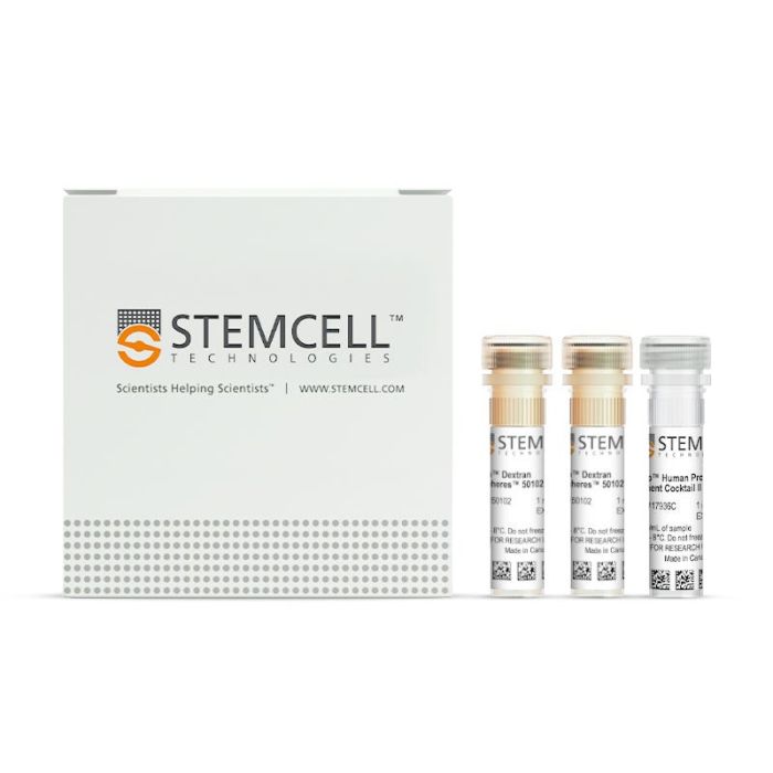
产品号 #17936_C
免疫磁珠负选试剂盒
免疫磁珠负选试剂盒
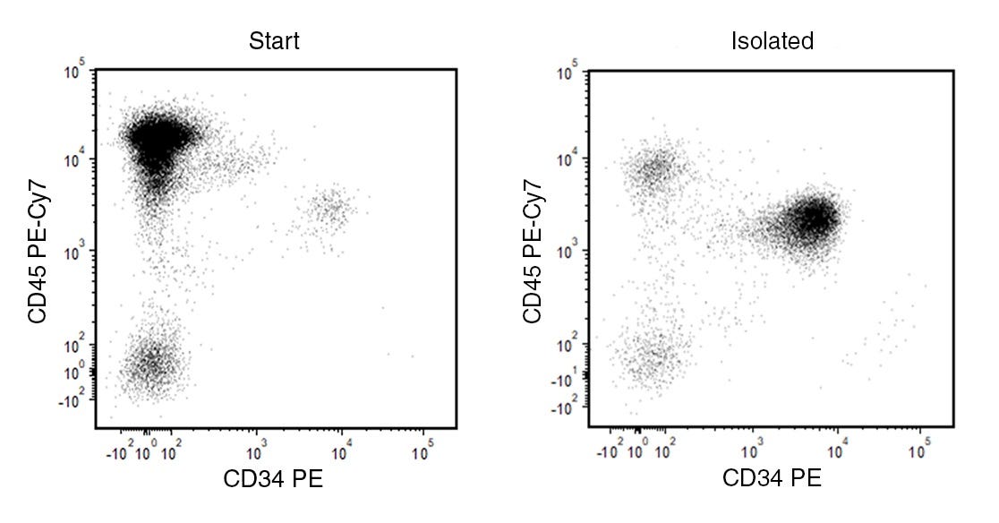
EasySep™ 人祖细胞富集试剂盒 II 旨在通过负选从新鲜脐血及其他细胞样本中分离造血祖细胞。操作流程包括通过抗体复合物和磁珠标记非目的细胞。通过EasySep™磁极将被磁珠标记的细胞与未被标记的目的细胞分离,接着简单地将目的细胞倾倒或吸至一个新的试管中。
磁体兼容性
• EasySep™ Magnet (Catalog #18000)
• “The Big Easy” EasySep™ Magnet (Catalog #18001)
• RoboSep™-S (Catalog #21000)
亚型
细胞分选试剂盒
细胞类型
造血干/祖细胞
种属
人
样本来源
Cord Blood
筛选方法
Negative
应用
细胞分选
品牌
EasySep,RoboSep
研究领域
干细胞生物学
Find supporting information and directions for use in the Product Information Sheet or explore additional protocols below.
This product is designed for use in the following research area(s) as part of the highlighted workflow stage(s). Explore these workflows to learn more about the other products we offer to support each research area.
| Species | Human |
|---|---|
| Magnet Compatibility | • EasySep™ Magnet (Catalog #18000) • “The Big Easy” EasySep™ Magnet (Catalog #18001) • RoboSep™-S (Catalog #21000) |
| Sample Source | Cord Blood |
| Selection Method | Negative |
通过免疫磁珠负选结合血小板去除技术分离未标记的人祖细胞
抗人CD45的小鼠单克隆IgG1抗体

小鼠单克隆IgG1抗体,抗人CD34
小鼠单克隆IgG1抗体,抗人、黑猩猩CD45
小鼠单克隆IgG1抗体,抗人CD34