产品号 #100-0020_C
Maturation kit for the generation of microglia from human ES and iPS cell-derived microglia precursors
For differentiation of human ES or iPS cells into hematopoietic progenitor cells
Differentiation kit for the generation of microglia precursors from human ES and iPS cell-derived hematopoietic progenitor cells
Dulbecco's Modified Eagle's Medium/Nutrient Ham's Mixture F-12 (DMEM/F-12) with 15 mM HEPES buffer
Sterile polypropylene conical tubes for use in cell centrifugation and other cell culture applications
Compatible antibodies for purity assessment of isolated cells
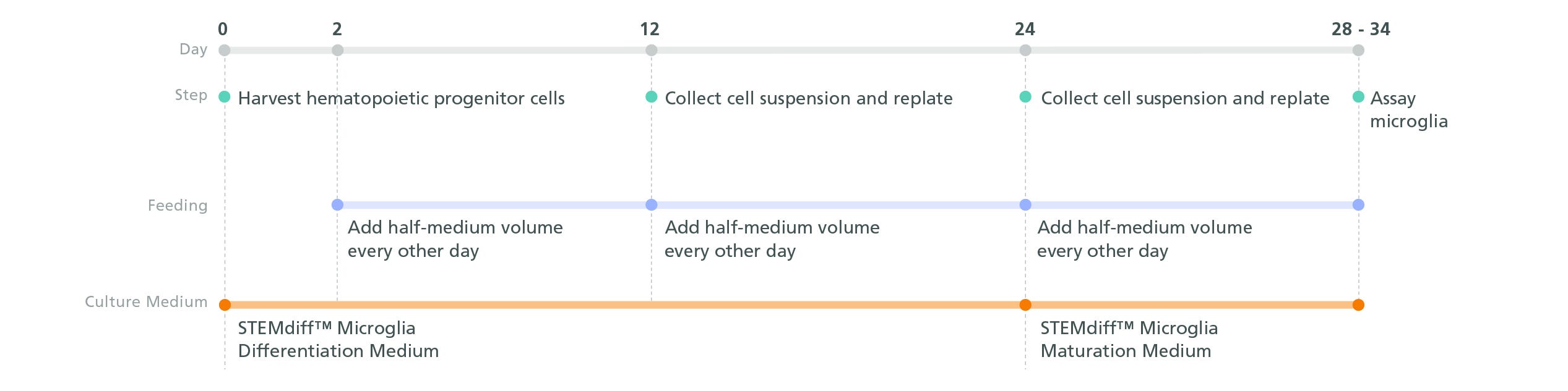
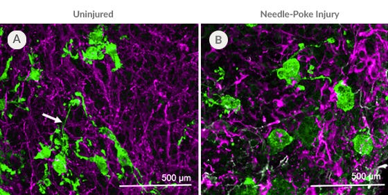
The STEMdiff™ Microglia Culture System comprises STEMdiff™ Microglia Differentiation Kit and STEMdiff™ Microglia Maturation Kit. Together, these kits are used to differentiate and mature microglia derived from human pluripotent stem cells (hPSCs) using STEMdiff™ Hematopoietic Kit (Catalog #05310).
Based on the protocol from the laboratory of Mathew Blurton-Jones (Abud et al., 2017), the resulting cells are a highly pure population of microglia (> 80% CD45/CD11b-positive, > 50% TREM2-positive microglia; < 20% morphologically distinct monocytes or macrophages).
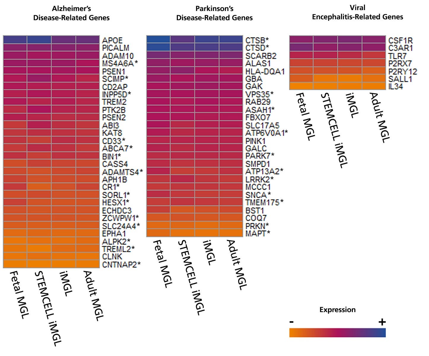
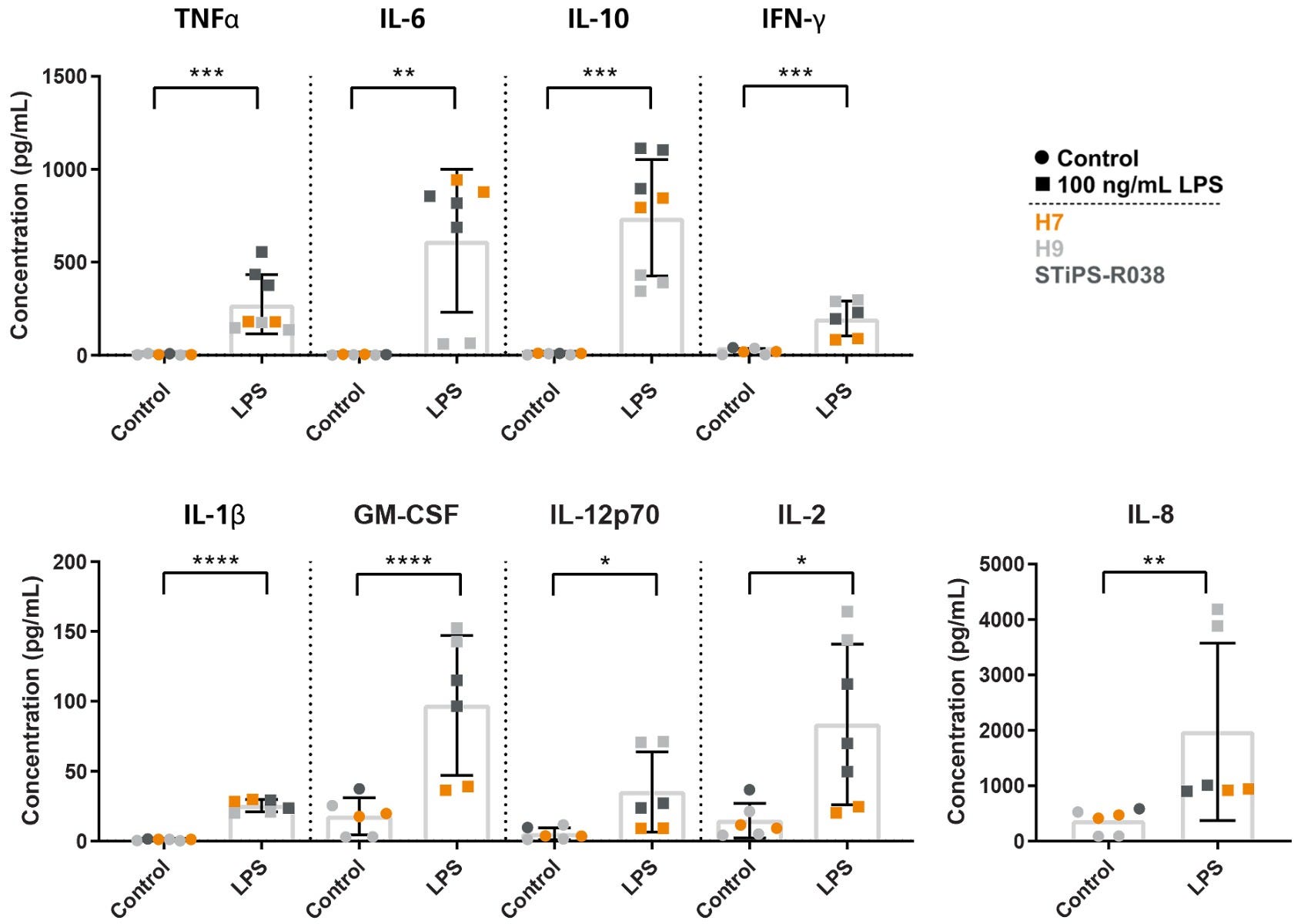
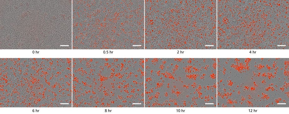
Cells derived using these products are versatile tools for modeling neuroinflammation, studying human neurological development and disease, co-culture applications, and toxicity testing.
Subtype
Specialized Media
Cell Type
Hematopoietic Cells, PSC-Derived, Microglia, Neural Cells, PSC-Derived
Species
Human
Application
Cell Culture, Differentiation
Brand
STEMdiff
Area of Interest
Disease Modeling, Drug Discovery and Toxicity Testing, Immunology, Neuroscience
Formulation Category
Serum-Free
Figure 1. Schematic for the STEMdiff™ Microglia Culture System Protocol
Functional microglia can be generated in 4-10 days from microglial precursors after 24 days in STEMdiff™ Microglia Differentiation Medium. For the differentiation of microglial precursors from hPSC-derived hematopoietic progenitor cells, see the PIS.
Figure 2. Microglia Generated with STEMdiff™ Microglia Culture System Express Disease-Relevant Genes Similar to Those from Published Differentiation and Maturation Protocols
Bulk RNA-seq datasets were extracted from 8 different publications that generated hPSC- (iMGL) and primary- (MGL) derived microglia and their transcriptional profiles compared to data from microglia generated with STEMdiff™ Microglia Culture System. The heat map displays absolute expression levels for select genes associated with Alzheimer’s disease, Parkinson’s disease, and viral encephalitis. Significant differences in gene expression between microglia generated with STEMdiff™ Microglia Culture System and any of the other 3 groups were identified by differential gene expression analysis. *= p<0.05 (DEseq2, adjusted). hPSC = human pluripotent stem cell.
Figure 3. Microglia Generated with STEMdiff™ Microglia Culture System Release Cytokines in Response to Inflammatory Signals
Microglia were generated using the STEMdiff™ Microglia Culture System and stimulated with 100 ng/mL LPS for 24 hours. The release of pro-inflammatory (TNFα, IL-6, IFN-γ, IL-1β, GM-CSF, IL-12p70, IL-2, IL-8) and anti-inflammatory (IL-10) cytokines were measured by MSD. The microglia release cytokines in response to LPS treatment, as expected. *, p ≤ 0.05; **, p ≤ 0.01; ***, p ≤ 0.001; ****, p ≤ 0.0001. LPS = lipopolysaccharide; MSD = Meso Scale Discovery.
Figure 4. STEMdiff™ Microglia Culture System Generates Functional Microglia Capable of Phagocytosis at Day 34
Microglia taking up pH-sensitive bioindicator particles at a concentration of 250 μg/mL (small dots) were measured over a 12-hour time period with live cell imaging. As the particles are phagocytosed, the cells turn red. Over time, the number of small dots decreased, and the red cells increased in number and aggregated. Scale bar = 100μm.
Figure 5. PSC-Derived Microglia Incorporate into Brain Organoids After 10 Days and Display an Activated Morphology upon Injury.
(A) Representative microglia and brain organoid co-cultures after 10 days, stained with IBA1 for microglia (green) and MAP2 for neurons (magenta). The microglia integrate among the neurons and display an unactivated morphology with extended processes (arrow). (B) The microglia display an activated amoeboid morphology upon injury as shown by IBA1 staining.
Find supporting information and directions for use in the Product Information Sheet or explore additional protocols below.
This product is designed for use in the following research area(s) as part of the highlighted workflow stage(s). Explore these workflows to learn more about the other products we offer to support each research area.
Thank you for your interest in IntestiCult™ Organoid Growth Medium (Human). Please provide us with your contact information and your local representative will contact you with a customized quote. Where appropriate, they can also assist you with a(n):
Estimated delivery time for your area
Product sample or exclusive offer
In-lab demonstration
扫描二维码或搜索微信号STEMCELLTech,即可关注我们的微信平台,第一时间接收丰富的技术资源和最新的活动信息。
如您有任何问题,欢迎发消息给STEMCELLTech微信公众平台,或与我们通过电话/邮件联系:400 885 9050 INFO.CN@STEMCELL.COM。