STEMdiff™小胶质细胞分化试剂盒

分化试剂盒用于从人胚胎干细胞和iPS细胞衍生的造血祖细胞生成小胶质细胞前体

产品号 #(选择产品)

产品号 #100-0019_C

分化试剂盒用于从人胚胎干细胞和iPS细胞衍生的造血祖细胞生成小胶质细胞前体

产品优势

  • 90%以上的源HPCs分化为小胶质细胞。
  • 与STEMdiff™造血试剂盒和STEMdiff™类神经器官共培养产生的HPCs上游兼容。
  • 允许分化为小胶质细胞,少量污染巨噬细胞或单核细胞。
  • 用该试剂盒生成的小胶质细胞表现出吞噬和活化。
  • 所需材料以简单,易于使用的格式提供。

产品组分包括

  • STEMdiff™小胶质细胞分化试剂盒(目录#100-0019)
    • STEMdiff™小胶质细胞基础培养基,90ml
    • STEMdiff™小胶质细胞补充剂1,10 mL
    • STEMdiff™Microglia Supplement, 2400 μL
Need a high-quality cell source? Use the hiPSC SCTi003-A (female) or SCTi004-A (male) control lines, manufactured with mTeSR™ Plus.
Products for Your Protocol
To see all required products for your protocol, please consult the Protocols and Documentation.

概述

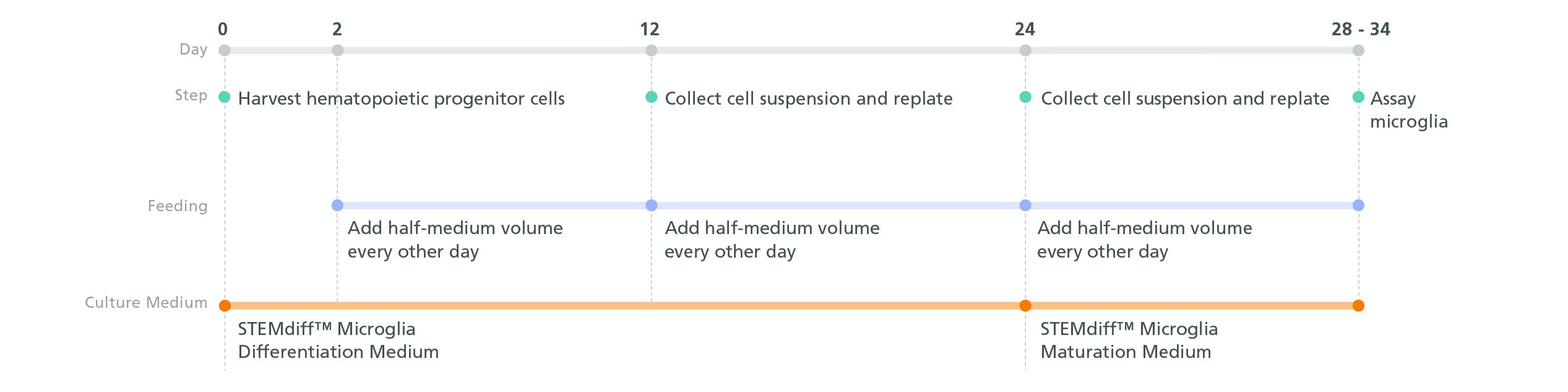
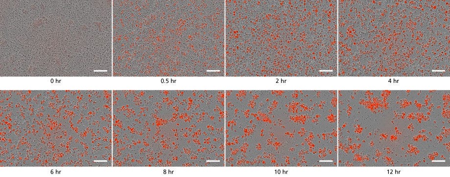
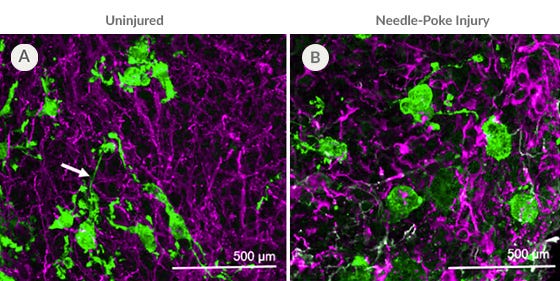
STEMdiff™小胶质细胞培养系统包括STEMdiff™小胶质细胞分化试剂盒和STEMdiff™小胶质细胞成熟试剂盒。使用STEMdiff™造血试剂盒(目录#05310),这些试剂盒可用于分化和成熟来自人类多能干细胞(hPSCs)的小胶质细胞。

根据Mathew Blurton-Jones (Abud et al., 2017)实验室的方案,得到的细胞是高度纯的小胶质细胞群(> 80% CD45/ cd11b阳性,> 50% trem2阳性;< 20%形态不同的单核细胞或巨噬细胞)。

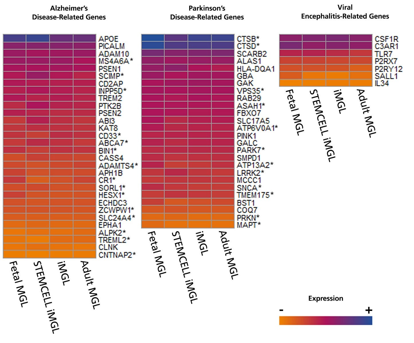
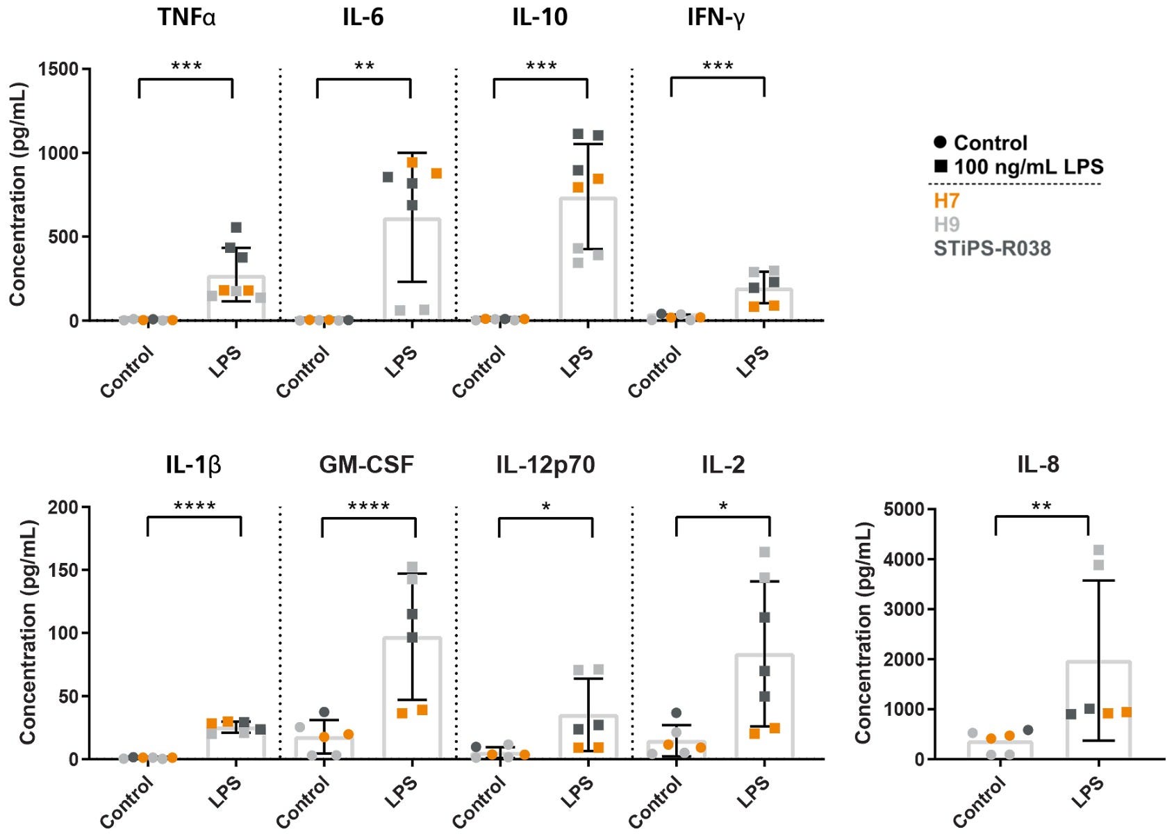
使用这些产品衍生的细胞是模拟神经炎症、研究人类神经发育和疾病、共培养应用和毒性测试的通用工具。

Subtype
Specialized Media
 
Cell Type
Hematopoietic Cells, PSC-Derived, Microglia, Neural Cells, PSC-Derived
 
Species
Human
 
Application
Cell Culture, Differentiation
 
Brand
STEMdiff
 
Area of Interest
Disease Modeling, Drug Discovery and Toxicity Testing, Immunology, Neuroscience
 
Formulation Category
Serum-Free
 

Data Figures

Experimental protocol schematic for generating microglial precursors from hPSC-derived hematopoietic progenitor cells.

Figure 1. Schematic for the STEMdiff™ Microglia Culture System Protocol

Microglial precursors can be generated in 24 days from hPSC-derived hematopoietic progenitor cells. For the generation of hematopoietic progenitor cells, see documentation for STEMdiff™ Hematopoietic Kit (Catalog #05310). For the maturation of microglial precursors to functional microglia, see the PIS.

Bulk RNA-seq heatmap showing that STEMdiff™ Microglia express disease-relevant genes similar to those from popular protocols.

Figure 2. Microglia Generated with STEMdiff™ Microglia Culture System Express Disease-Relevant Genes Similar to Those from Published Differentiation and Maturation Protocols

Bulk RNA-seq datasets were extracted from 8 different publications that generated hPSC- (iMGL) and primary- (MGL) derived microglia and their transcriptional profiles compared to data from microglia generated with STEMdiff™ Microglia Culture System. The heat map displays absolute expression levels for select genes associated with Alzheimer’s disease, Parkinson’s disease, and viral encephalitis. Significant differences in gene expression between microglia generated with STEMdiff™ Microglia Culture System and any of the other 3 groups were identified by differential gene expression analysis. *= p<0.05 (DEseq2, adjusted). hPSC = human pluripotent stem cell.

Graphs demonstrating that STEMdiff™ Microglia release cytokines in response to LPS stimulation, compared to control.

Figure 3. Microglia Generated with STEMdiff™ Microglia Culture System Release Cytokines in Response to Inflammatory Signals

Microglia were generated using the STEMdiff™ Microglia Culture System and stimulated with 100 ng/mL LPS for 24 hours. The release of pro-inflammatory (TNFα, IL-6, IFN-γ, IL-1β, GM-CSF, IL-12p70, IL-2, IL-8) and anti-inflammatory (IL-10) cytokines were measured by MSD. The microglia release cytokines in response to LPS treatment, as expected. *, p ≤ 0.05; **, p ≤ 0.01; ***, p ≤ 0.001; ****, p ≤ 0.0001. LPS = lipopolysaccharide; MSD = Meso Scale Discovery.

Time lapse of functional microglia phagocytosing bioindicator particles in culture. Over time, cells turn red and aggregate.

Figure 4. STEMdiff™ Microglia Culture System Generates Functional Microglia Capable of Phagocytosis at Day 34

Microglia taking up pH-sensitive bioindicator particles at a concentration of 250 μg/mL (small dots) were measured over a 12-hour time period with live cell imaging. As the particles are phagocytosed, the cells turn red. Over time, the number of small dots decreased, and the red cells increased in number and aggregated. Scale bar = 100μm.

Figure 5. PSC-Derived Microglia Incorporate into Brain Organoids After 10 Days and Display an Activated Morphology upon Injury.

(A) Representative microglia and brain organoid co-cultures after 10 days, stained with IBA1 for microglia (green) and MAP2 for neurons (magenta). The microglia integrate among the neurons and display an unactivated morphology with extended processes (arrow). (B) The microglia display an activated amoeboid morphology upon injury as shown by IBA1 staining.

Protocols and Documentation

Find supporting information and directions for use in the Product Information Sheet or explore additional protocols below.

Document Type
Product Name
Catalog #
Lot #
Language
Catalog #
100-0019
Lot #
All
Language
English
Document Type
Safety Data Sheet 1
Catalog #
100-0019
Lot #
All
Language
English
Document Type
Safety Data Sheet 2
Catalog #
100-0019
Lot #
All
Language
English
Document Type
Safety Data Sheet 3
Catalog #
100-0019
Lot #
All
Language
English

Applications

This product is designed for use in the following research area(s) as part of the highlighted workflow stage(s). Explore these workflows to learn more about the other products we offer to support each research area.

Resources and Publications

Educational Materials (27)

更多信息

更多信息
Species Human
Formulation Category Serum-Free
Legal Statement: Copyright © 2024 by STEMCELL Technologies Inc. All rights reserved including graphics and images. STEMCELL Technologies & Design, STEMCELL Shield Design, Scientists Helping Scientists, and STEMdiff are trademarks of STEMCELL Technologies Canada Inc. All other trademarks are the property of their respective holders. While STEMCELL has made all reasonable efforts to ensure that the information provided by STEMCELL and its suppliers is correct, it makes no warranties or representations as to the accuracy or completeness of such information. PRODUCTS ARE FOR RESEARCH USE ONLY AND NOT INTENDED FOR HUMAN OR ANIMAL DIAGNOSTIC OR THERAPEUTIC USES UNLESS OTHERWISE STATED. FOR ADDITIONAL INFORMATION ON QUALITY AT STEMCELL, REFER TO WWW.STEMCELL.COM/COMPLIANCE.
版权 © 2025 STEMCELL Technologies 技术有限公司。保留所有权利。