STEMdiff™ 脑类器官试剂盒

用于人脑类器官建立与成熟的培养基试剂盒

产品号 #(选择产品)

产品号 #08570_C

用于人脑类器官建立与成熟的培养基试剂盒

产品组分包括

  •  STEMdiff™ 脑类器官基础培养基 1,100 mL
  • STEMdiff™ 脑类器官基础培养基 2,250 mL
  • STEMdiff™ 脑类器官补充剂 A,10 mL
  •  STEMdiff™ 脑类器官补充剂 B,0.5 mL
  • STEMdiff™ 脑类器官补充剂 C,0.25 mL
  •  STEMdiff™ 脑类器官补充剂 D,0.5 mL
  •  STEMdiff™ 脑类器官补充剂 E,4.5 mL
Need a high-quality cell source? Use the hiPSC SCTi003-A (female) control line, manufactured with mTeSR™ Plus.

What Our Scientist Says

Human brain development is complex, so to observe it in a dish is a huge scientific breakthrough. We've tried to simplify that process and make it more accessible to you, regardless of how much stem cell experience you have.

Leon ChewScientist
Leon Chew, Scientist

总览

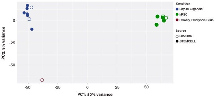
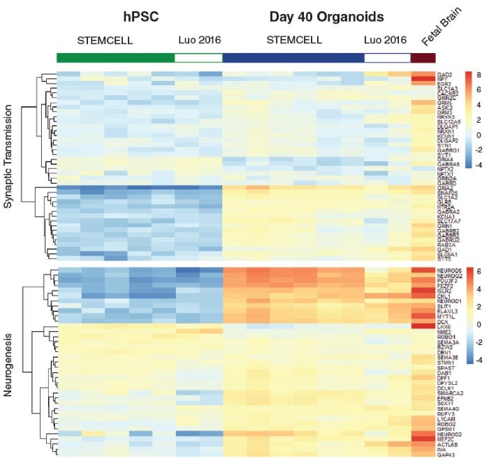
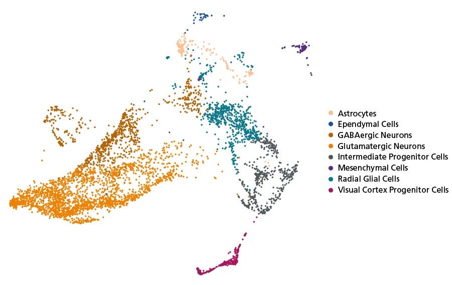
生成自组织的、由多能干细胞 (PSC) 衍生的神经类器官,其细胞组成和结构组织与发育中的人脑相似。

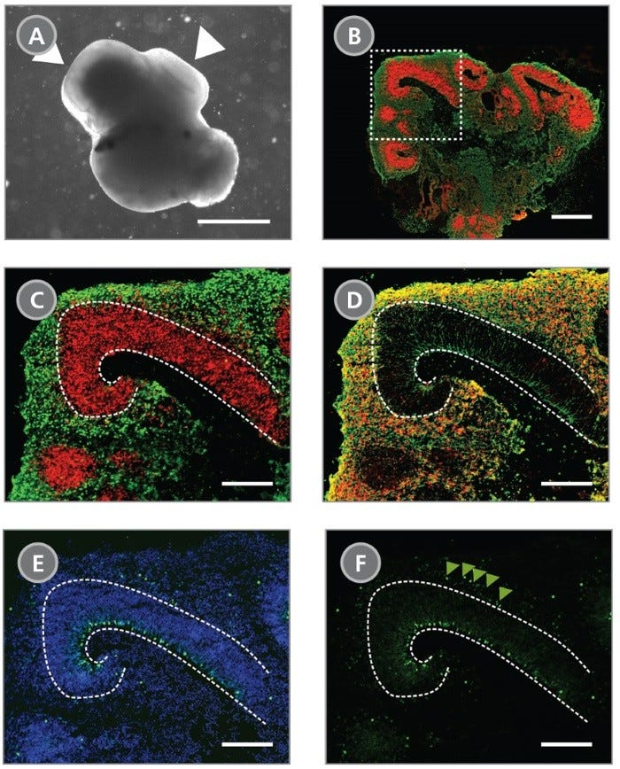
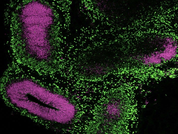
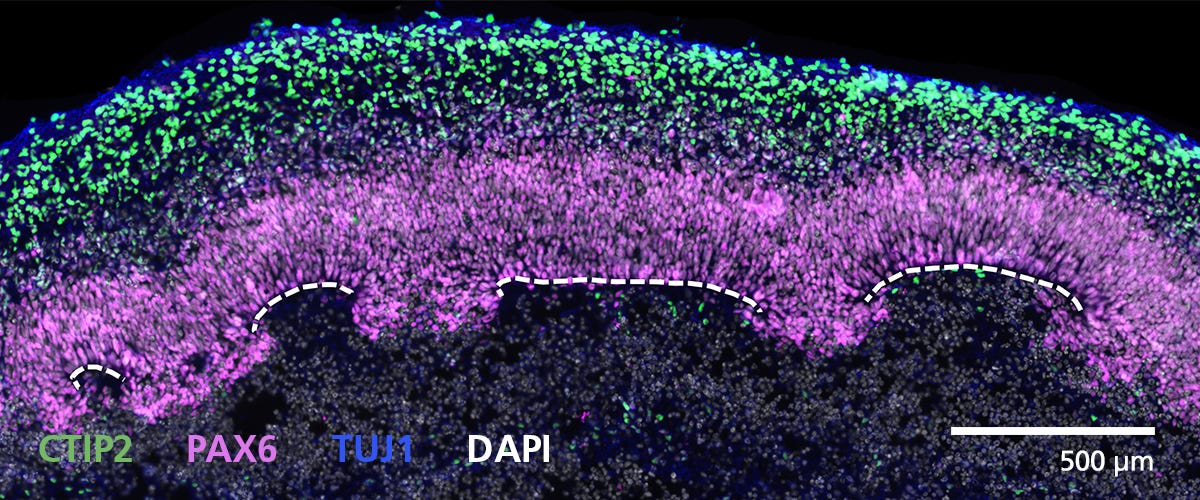
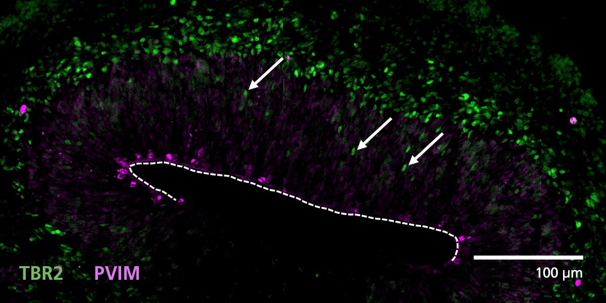
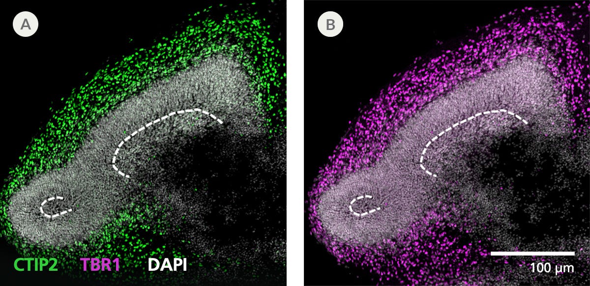
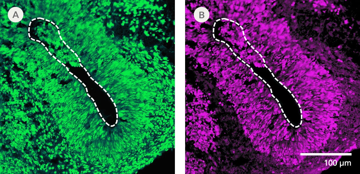
这些成分明确的无血清细胞培养基和简单的四阶段实验方案基于 Lancaster 等人发表的配方(Lancaster MA 等人,Nature,2013 和 Lancaster MA 等人,Science,2014),旨在更可靠地生成脑类器官。使用 STEMdiff™ 脑类器官试剂盒生成的类器官从胚状体 (EB) 形成步骤开始,随后神经上皮细胞扩增,具有皮质样区域,包括脑室区 (PAX6+/SOX2+/Ki-67+)、外脑室下区 (Ki-67+/p-Vimentin+)、中间区 (TBR2+) 和皮质板 (CTIP2+/MAP2+/TBR1+),其分层方向与体内观察到的相似。

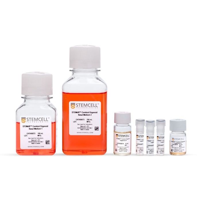
对于长期培养(> 40 天),成熟所需的成分可作为STEMdiff™ 脑类器官成熟试剂盒购买。

亚型
专用培养基
 
细胞类型
神经细胞,PSC衍生,神经干/祖细胞,多能干细胞
 
种属

 
应用
细胞培养,鉴定,分化,功能学筛选,免疫荧光,类器官培养,表型鉴定,球状体培养
 
品牌
STEMdiff
 
研究领域
疾病建模,药物发现和毒理检测,神经科学,干细胞生物学
 
制剂类别
无血清
 

Data Figures

Figure 1. Cerebral Organoids Contain Multiple Layered Regions That Recapitulate the Cortical Lamination Process Observed During In Vivo Human Brain Development

(A) A representative phase-contrast image of a whole cerebral organoid at Day 40 generated using the STEMdiff™ Cerebral Organoid Kit. Cerebral organoids at this stage are made up of phase-dark structures that may be surrounded by regions of thinner, more translucent structures that display layering (arrowheads). (B) Immunohistological analysis on cryosections of cerebral organoids reveals cortical regions within the organoid labeled by the apical progenitor marker PAX6 (red) and neuronal marker β-tubulin III+ (TUJ-1) (green). (C-F) Inset of boxed region from (B). (C) PAX6+ apical progenitors (red, enclosed by dotted line) are localized to a ventricular zone-like region. β-tubulin III+ neurons (green) are adjacent to the ventricular zone. (D) CTIP2, a marker of the developing cortical plate, co-localizes with β-tubulin III+ neurons in a cortical plate-like region. Organization of the layers recapitulates early corticogenesis observed during human brain development. (E) Proliferating progenitor cells labeled by Ki-67 (green) localize along the ventricle, nuclei are counterstained with DAPI (blue). (F) An additional population of Ki-67+ cells is found in an outer subventricular zone-like region (arrowheads).

Figure 2. Cerebral Organoids Generated with the STEMdiff™ Cerebral Organoid Kit Are Transcriptionally Similar to Those from Published Protocols

RNA-seq data was extracted from a publication (C Luo et al. Cell Rep, 2016) that generated cerebral organoids (open blue circles) and compared this transcriptional profile to that of cerebral organoids generated with the STEMdiff™ Cerebral Organoid Kit (filled blue circles). Principal component analysis was performed on these data. The cerebral organoids from the STEMdiff™ Cerebral Organoid Kit cluster together, and cluster with the previously published cerebral organoids. The first principal component accounts for the majority of variance seen (PC1; 80%) and distinguishes the cerebral organoid samples from the hPSCs (green circles). The second principal component accounts for only 9% of the variation, and highlights the modest expression differences between cultured organoids and primary embryonic fetal brain samples (19 post-conceptional weeks, brown circles).

Figure 3. Neural Organoids Generated with STEMdiff™ Cerebral Organoid Kit Express Expected Key Markers

RNA from cerebral organoids generated with the STEMdiff™ Cerebral Organoid Kit was harvested and assayed using bulk RNA-seq. A heatmap of expression levels for genes associated with synaptic transmission function and neurogenesis in Day 40 organoids demonstrate that gene expression of cerebral organoids generated from the STEMdiff™ Cerebral Organoid Kit are similar to published results (C Luo et al. Cell Rep, 2016).

Immunocytochemistry image of a cerebral organoid cultured in mTeSR™ Plus and directed to cerebral organoids using the STEMdiff™ Cerebral Organoid Kit.

Figure 4. Cerebral Organoids Can Be Generated from hPSCs Maintained in mTeSR™ Plus

Human ES (H9) cells were cultured with mTeSR™ Plus and directed to cerebral organoids using the STEMdiff™ Cerebral Organoid Kit. Image shows apical progenitor marker SOX2 (magenta) and neuronal marker TBR1 (green).

Figure 5. Cryosectioned Cerebral Organoids Show Stratification of Cortical Plate Neurons and Progenitor Zones

Cerebral organoids were generated using the STEMdiff™ Cerebral Organoid Kit. A 16-μm-thick section of a Day 40 cerebral organoid was stained for CTIP2 (green), PAX6 (magenta), βIII-tubulin/TUJ1 (blue), and DAPI (gray). Cortical regions are defined by progenitor cells (PAX6+) that are radially organized around a pseudo-ventricle (dashed line). These progenitors give rise to cortical plate neurons indicated by CTIP2 and TUJ1 expression. For a detailed cryogenic tissue processing and immunofluorescence protocol, please see the Methods Library.

Figure 6. Zones of Active Proliferation in Cerebral Organoids Are Preserved Following the Protocol for Tissue Processing

Organoid tissue was processed for immunofluorescence and stained for TBR2 (intermediate precursors, green) and phosphorylated vimentin (PVIM, dividing cells, magenta). Cells actively divide at the apical border of cortical regions along the border of the pseudo-ventricle (dashed line). A population of these dividing cells will express TBR2 and then migrate (arrows) from the progenitor zone to form a layer of intermediate progenitors. For a detailed cryogenic tissue processing and immunofluorescence protocol, please see the Methods Library.

Figure 7. Immunofluorescence from Cryosectioned Cerebral Organoids Indicates Preserved Organization of Cortical Neurons

Organoid tissue was processed for immunofluorescence and stained for CTIP2 (green), TBR1 (layer 5/6 cortical neurons, magenta), and DAPI (white). Deep layer neuronal markers CTIP2 and TBR1 are expressed in cells around presumptive progenitor zones (dashed line) toward the outside or apical surface of organoids. For a detailed cryogenic tissue processing and immunofluorescence protocol, please see the Methods Library.

Figure 8. Cryogenic Tissue Processing and Immunofluorescence Captures Arrangement of Neural Progenitors Around Pseudo-Ventricles in Cerebral Organoids

Organoid tissue was processed for immunofluorescence and stained for (A) FOXG1 (forebrain cells, green) or (B) SOX2 (neural progenitors, magenta). Organoids derived from STEMdiff™ Cerebral Organoid Kit generate forebrain-type tissue as indicated by FOXG1 expression. Neural progenitors expressing SOX2 are radially arranged around a pseudo-ventricle area (dashed line). For a detailed cryogenic tissue processing and immunofluorescence protocol, please see the Methods Library.

scRNA seq data for STEMdiff™ Cerebral Organoid Kit

Figure 9. Neural Organoids Generated with STEMdiff™ Cerebral Organoid Kit Contain Diverse Cell Types

Unguided cerebral organoids were generated from H9 ESCs using STEMdiff™ Cerebral Organoid Kit. scRNA-seq gene expression data captures the cellular diversity of these neural organoids which can be further explored using the Single Cell RNA Sequencing Data Visualization Tool for Neural Organoids. At Day 50, the organoids were dissociated into a single-cell suspension. The library was prepared using Chromium Single Cell 3ʹ v1 protocol with Feature Barcoding technology (10x Genomics) following surface protein staining with TotalSeq™–B (BioLegend). The barcoded processing, gene counting, and aggregation were done using the Cell Ranger software v3.1.0. Further processing and demultiplexing was done with Seurat v4.1.1. The data have been made publicly available on GEO: GSE218457. ESC = embryonic stem cell

Protocols and Documentation

Find supporting information and directions for use in the Product Information Sheet or explore additional protocols below.

Document Type
Product Name
Catalog #
Lot #
Language
Catalog #
08570
Lot #
All
Language
English
Document Type
Safety Data Sheet 1
Catalog #
08570
Lot #
All
Language
English
Document Type
Safety Data Sheet 2
Catalog #
08570
Lot #
All
Language
English
Document Type
Safety Data Sheet 3
Catalog #
08570
Lot #
All
Language
English
Document Type
Safety Data Sheet 4
Catalog #
08570
Lot #
All
Language
English
Document Type
Safety Data Sheet 5
Catalog #
08570
Lot #
All
Language
English
Document Type
Safety Data Sheet 6
Catalog #
08570
Lot #
All
Language
English
Document Type
Safety Data Sheet 7
Catalog #
08570
Lot #
All
Language
English

Applications

This product is designed for use in the following research area(s) as part of the highlighted workflow stage(s). Explore these workflows to learn more about the other products we offer to support each research area.

Resources and Publications

Educational Materials (46)

Brochure

Publications (5)

Human CNS barrier-forming organoids with cerebrospinal fluid production. L. Pellegrini et al. Science (New York, N.Y.) 2020

Abstract

Cerebrospinal fluid (CSF) is a vital liquid, providing nutrients and signaling molecules and clearing out toxic by-products from the brain. The CSF is produced by the choroid plexus (ChP), a protective epithelial barrier that also prevents free entry of toxic molecules or drugs from the blood. Here, we establish human ChP organoids with a selective barrier and CSF-like fluid secretion in self-contained compartments. We show that this in vitro barrier exhibits the same selectivity to small molecules as the ChP in vivo and that ChP-CSF organoids can predict central nervous system (CNS) permeability of new compounds. The transcriptomic and proteomic signatures of ChP-CSF organoids reveal a high degree of similarity to the ChP in vivo. Finally, the intersection of single-cell transcriptomics and proteomic analysis uncovers key human CSF components produced by previously unidentified specialized epithelial subtypes.
One-Stop Microfluidic Assembly of Human Brain Organoids To Model Prenatal Cannabis Exposure. Z. Ao et al. Analytical chemistry 2020

Abstract

Prenatal cannabis exposure (PCE) influences human brain development, but it is challenging to model PCE using animals and current cell culture techniques. Here, we developed a one-stop microfluidic platform to assemble and culture human cerebral organoids from human embryonic stem cells (hESC) to investigate the effect of PCE on early human brain development. By incorporating perfusable culture chambers, air-liquid interface, and one-stop protocol, this microfluidic platform can simplify the fabrication procedure and produce a large number of organoids (169 organoids per 3.5 cm × 3.5 cm device area) without fusion, as compared with conventional fabrication methods. These one-stop microfluidic assembled cerebral organoids not only recapitulate early human brain structure, biology, and electrophysiology but also have minimal size variation and hypoxia. Under on-chip exposure to the psychoactive cannabinoid, $\Delta$-9-tetrahydrocannabinol (THC), cerebral organoids exhibited reduced neuronal maturation, downregulation of cannabinoid receptor type 1 (CB1) receptors, and impaired neurite outgrowth. Moreover, transient on-chip THC treatment also decreased spontaneous firing in these organoids. This one-stop microfluidic technique enables a simple, scalable, and repeatable organoid culture method that can be used not only for human brain organoids but also for many other human organoids including liver, kidney, retina, and tumor organoids. This technology could be widely used in modeling brain and other organ development, developmental disorders, developmental pharmacology and toxicology, and drug screening.
Cerebral organoids at the air–liquid interface generate diverse nerve tracts with functional output S. L. Giandomenico et al. Nature Neuroscience 2019 apr

Abstract

Neural organoids have the potential to improve our understanding of human brain development and neurological disorders. However, it remains to be seen whether these tissues can model circuit formation with functional neuronal output. Here we have adapted air–liquid interface culture to cerebral organoids, leading to improved neuronal survival and axon outgrowth. The resulting thick axon tracts display various morphologies, including long-range projection within and away from the organoid, growth-cone turning, and decussation. Single-cell RNA sequencing reveals various cortical neuronal identities, and retrograde tracing demonstrates tract morphologies that match proper molecular identities. These cultures exhibit active neuronal networks, and subcortical projecting tracts can innervate mouse spinal cord explants and evoke contractions of adjacent muscle in a manner dependent on intact organoid-derived innervating tracts. Overall, these results reveal a remarkable self-organization of corticofugal and callosal tracts with a functional output, providing new opportunities to examine relevant aspects of human CNS development and disease.

更多信息

更多信息
Species Human
Formulation Category Serum-Free
Legal Statement: This product was developed under a license to intellectual property owned by the Institute of Molecular Biotechnology (IMBA) of the Austrian Academy of Sciences. This product is sold for research use only (whether the buyer is an academic or for-profit entity) under a non-transferable, limited-use license. Purchase of this product does not include the right to sell, use or otherwise transfer this product for commercial purposes (i.e., any activity undertaken for consideration, such as use of this product for manufacturing, or resale of this product or any materials made using this product, or use of this product or any materials made using this product to provide services or, in collaboration with, a for-profit entity, for purposes other than research applications (i.e., drug development activities). Purchasers wishing to use the product for commercial purposes should contact IMBA at technologvtransfer@imba.oeaw.ac.at. PRODUCTS ARE FOR RESEARCH USE ONLY AND NOT INTENDED FOR HUMAN OR ANIMAL DIAGNOSTIC OR THERAPEUTIC USES UNLESS OTHERWISE STATED. FOR ADDITIONAL INFORMATION ON QUALITY AT STEMCELL, REFER TO WWW.STEMCELL.COM/COMPLIANCE.
版权 © 2025 STEMCELL Technologies 技术有限公司。保留所有权利。