RosetteSep™人CD45去除抗体混合物

免疫密度负选试剂混合物

产品号 #(选择产品)

产品号 #15122_C

免疫密度负选试剂混合物

产品优势

  • 快捷、操作简单
  • 不需要特殊设备或额外培训
  • 获得的活细胞无标记
  • 可与SepMate™联合使用,实现一致的高通量样本处理

产品组分包括

  • RosetteSep™人CD45细胞去除抗体混合物(产品号 #15624)
    • RosetteSep™人CD45去除抗体混合物,2mL
  • RosetteSep™人粒细胞去除抗体混合物(产品号 #15624)
    • RosetteSep™人CD45去除抗体混合物 , 5x2mL
New look, same high quality and support! You may notice that your instrument or reagent packaging looks slightly different from images displayed on the website, or from previous orders. We are updating our look but rest assured, the products themselves and how you should use them have not changed. Learn more

总览

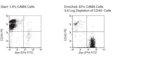
RosetteSep™人CD45去除抗体混合物通过去除CD45+细胞从全血中富集上皮循环肿瘤细胞(CTC)。四聚体抗体复合物可识别CD45 、CD66b以及红细胞(RBC)上的糖蛋白A,从而靶向去除非目的细胞。使用密度梯度离心液如Lymphoprep™(产品号 #18060)离心后 ,非目的细胞会与红细胞一起沉淀。纯化的上皮肿瘤细胞为血浆和密度梯度离心液的交界界面中高度富集的细胞。

亚型
细胞分选试剂盒
 
细胞类型
癌细胞及细胞系
 
种属

 
样本来源
Buffy Coat,Whole Blood
 
筛选方法
删除
 
应用
细胞分选
 
品牌
RosetteSep
 
研究领域
癌症,免疫,干细胞生物学
 

Data Figures

FACS Profile Results with RosetteSep™ Human CD45+ Cell Depletion Cocktail

Figure 1. FACS Profile Results with RosetteSep™ Human CD45+ Cell Depletion Cocktail

Ber-EP4 is an antibody against an epithelial cell surface antigen.

Protocols and Documentation

Find supporting information and directions for use in the Product Information Sheet or explore additional protocols below.

Document Type
Product Name
Catalog #
Lot #
Language
Catalog #
15122, 15162
Lot #
All
Language
English
Document Type
Safety Data Sheet
Catalog #
15122, 15162
Lot #
All
Language
English

Resources and Publications

Educational Materials (4)

Frequently Asked Questions

What is RosetteSep™?

RosetteSep™ is a rapid cell separation procedure for the isolation of purified cells directly from whole blood, without columns or magnets.

How does RosetteSep™ work?

The antibody cocktail crosslinks unwanted cells to red blood cells (RBCs), forming rosettes. The unwanted cells then pellet with the free RBCs when centrifuged over a density centrifugation medium (e.g. Ficoll-Paque™ PLUS, Lymphoprep™).

What factors affect cell recovery?

The temperature of the reagents can affect cell recovery. All reagents should be at room temperature (sample, density centrifugation medium, PBS, centrifuge) before performing the isolations. Layering can also affect recovery so be sure to carefully layer the sample to avoid mixing with the density centrifugation medium as much as possible. Be sure to collect the entire enriched culture without disturbing the RBC pellet. A small amount of density centrifugation medium can be collected without worry.

Which cell samples can RosetteSep™ be used with?

RosetteSep™ can be used with leukapheresis samples, bone marrow or buffy coat, as long as: the concentration of cells does not exceed 5 x 10⁷ per mL (can dilute if necessary); and there are at least 100 RBCs for every nucleated cell (RBCs can be added if necessary).

Can RosetteSep™ be used with previously frozen or cultured cells?

Yes. Cells should be re-suspended at 2 - 5 x 10⁷ cells / mL in PBS + 2% FBS. Fresh whole blood should be added at 250 µL per mL of sample, as a source of red cells.

Can RosetteSep™ be used to enrich progenitors from cord blood?

Yes. Sometimes cord blood contains immature nucleated red cells that have a lower density than mature RBCs. These immature red cells do not pellet over Ficoll™, which can lead to a higher RBC contamination than peripheral blood separations.

Does RosetteSep™ work with mouse cells?

No, but we have developed EasySep™, a magnetic-based cell isolation system which works with mouse and other non-human species.

Which anticoagulant should be used with RosetteSep™?

Peripheral blood should be collected in heparinized Vacutainers. Cord blood should be collected in ACD.

Should the anticoagulant be washed off before using RosetteSep™?

No, the antibody cocktail can be added directly to the sample.

Publications (12)

Sequential Circulating Tumor Cell Counts in Patients with Locally Advanced or Metastatic Hepatocellular Carcinoma: Monitoring the Treatment Response. K.-M. Rau et al. Journal of clinical medicine 2020 jan

Abstract

Hepatocellular carcinoma (HCC) is among the most common causes of cancer death in men. Whether or not a longitudinal follow-up of circulating tumor cells (CTCs) before and at different time points during systemic/targeted therapy is useful for monitoring the treatment response of patients with locally advanced or metastatic HCC has been evaluated in this study. Blood samples (n = 104) were obtained from patients with locally advanced or metastatic HCC (n = 30) for the enrichment of CTCs by a negative selection method. Analysis of the blood samples from patients with defined disease status (n = 81) revealed that those with progressive disease (PD, n = 37) had significantly higher CTC counts compared to those with a partial response (PR) or stable disease (SD; n = 44 for PR + SD, p = 0.0002). The median CTC count for patients with PD and for patients with PR and SD was 50 (interquartile range 21-139) and 15 (interquartile range 4-41) cells/mL of blood, respectively. A longitudinal analysis of patients (n = 17) after a series of blood collections demonstrated that a change in the CTC count correlated with the patient treatment response in most of the cases and was particularly useful for monitoring patients without elevated serum alpha-fetoprotein (AFP) levels. Sequential CTC enumeration during treatment can supplement standard medical tests and benefit the management of patients with locally advanced or metastatic HCC, in particular for the AFP-low cases.
Sensitive and easy screening for circulating tumor cells by flow cytometry. A. Lopresti et al. JCI insight 2019 jun

Abstract

Circulating Tumor Cells (CTCs) represent an easy, repeatable and representative access to information regarding solid tumors. However, their detection remains difficult because of their paucity, their short half-life, and the lack of reliable surface biomarkers. Flow cytometry (FC) is a fast, sensitive and affordable technique, ideal for rare cells detection. Adapted to CTCs detection (i.e. extremely rare cells), most FC-based techniques require a time-consuming pre-enrichment step, followed by a 2-hours staining procedure, impeding on the efficiency of CTCs detection. We overcame these caveats and reduced the procedure to less than one hour, with minimal manipulation. First, cells were simultaneously fixed, permeabilized, then stained. Second, using low-speed FC acquisition conditions and two discriminators (cell size and pan-cytokeratin expression), we suppressed the pre-enrichment step. Applied to blood from donors with or without known malignant diseases, this protocol ensures a high recovery of the cells of interest independently of their epithelial-mesenchymal plasticity and can predict which samples are derived from cancer donors. This proof-of-concept study lays the bases of a sensitive tool to detect CTCs from a small amount of blood upstream of in-depth analyses.
Application of Prostate Cancer Models for Preclinical Study: Advantages and Limitations of Cell Lines, Patient-Derived Xenografts, and Three-Dimensional Culture of Patient-Derived Cells. T. Namekawa et al. Cells 2019 jan

Abstract

Various preclinical models have been developed to clarify the pathophysiology of prostate cancer (PCa). Traditional PCa cell lines from clinical metastatic lesions, as exemplified by DU-145, PC-3, and LNCaP cells, are useful tools to define mechanisms underlying tumorigenesis and drug resistance. Cell line-based experiments, however, have limitations for preclinical studies because those cells are basically adapted to 2-dimensional monolayer culture conditions, in which the majority of primary PCa cells cannot survive. Recent tissue engineering enables generation of PCa patient-derived xenografts (PDXs) from both primary and metastatic lesions. Compared with fresh PCa tissue transplantation in athymic mice, co-injection of PCa tissues with extracellular matrix in highly immunodeficient mice has remarkably improved the success rate of PDX generation. PDX models have advantages to appropriately recapitulate the molecular diversity, cellular heterogeneity, and histology of original patient tumors. In contrast to PDX models, patient-derived organoid and spheroid PCa models in 3-dimensional culture are more feasible tools for in vitro studies for retaining the characteristics of patient tumors. In this article, we review PCa preclinical model cell lines and their sublines, PDXs, and patient-derived organoid and spheroid models. These PCa models will be applied to the development of new strategies for cancer precision medicine.

更多信息

更多信息
Species Human
Sample Source Buffy Coat, Whole Blood
Selection Method Depletion
PRODUCTS ARE FOR RESEARCH USE ONLY AND NOT INTENDED FOR HUMAN OR ANIMAL DIAGNOSTIC OR THERAPEUTIC USES UNLESS OTHERWISE STATED. FOR ADDITIONAL INFORMATION ON QUALITY AT STEMCELL, REFER TO WWW.STEMCELL.COM/COMPLIANCE.
版权 © 2025 STEMCELL Technologies 技术有限公司。保留所有权利。