STEMdiff™SMADi神经诱导试剂盒

高效SMAD抑制介导的人ES和iPS细胞神经诱导无血清培养基试剂盒

产品号 #(选择产品)

产品号 #08581_C

高效SMAD抑制介导的人ES和iPS细胞神经诱导无血清培养基试剂盒

产品优势

  • 明确且无血清
  • 促进ES和iPS细胞向cns型npc的有效转化,并抑制非cns细胞类型的不必要分化
  • 高效神经诱导,甚至难以分化的胚胎干细胞和iPS细胞系
  • 提高下游分化为神经元和胶质细胞的效率
  • 兼容胚状体和单层培养协议的神经诱导
  • 使在任何TeSR™家族维持培养基中维持的细胞系可重复分化
  • 方便,用户友好的格式和协议
Need a high-quality cell source? Use the hiPSC SCTi003-A (female) or SCTi004-A (male) control lines, manufactured with mTeSR™ Plus.

概述

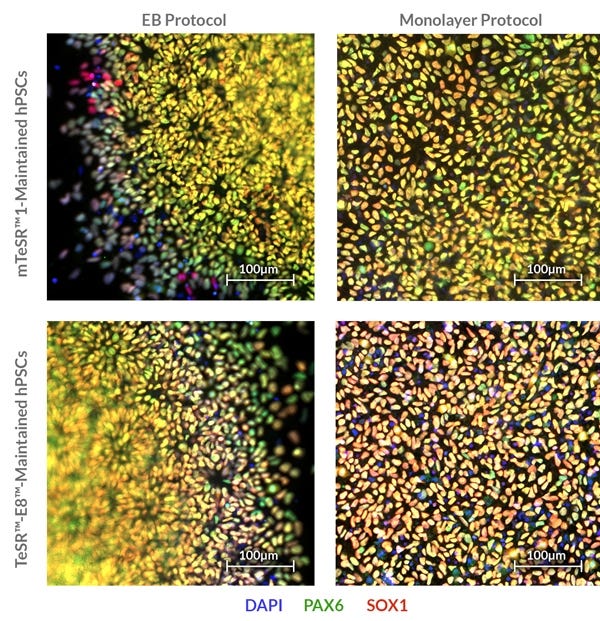
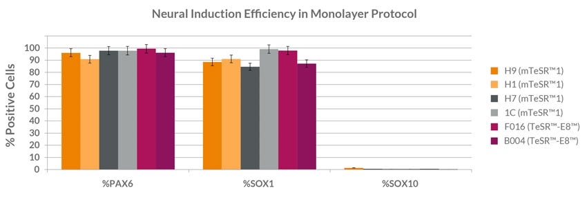
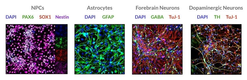
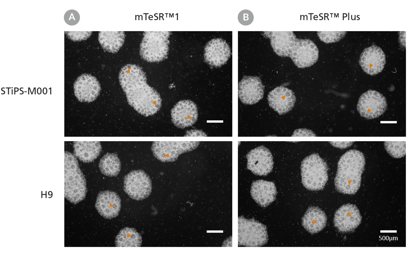
STEMdiff™SMADi神经诱导试剂盒由定义的无血清培养基和补充物组成,用于高效神经诱导人胚胎干细胞(ES)和诱导多能干细胞(iPS)细胞。该试剂盒结合STEMdiff™神经诱导培养基(目录#05835)和STEMdiff™SMADi神经诱导补充剂,通过阻断TGF-β/ bmp依赖的SMAD信号传导来指导分化,从而有效地神经诱导甚至难以分化的细胞系。神经祖细胞(npc)可以使用STEMdiff™SMADi神经诱导试剂盒与胚状体(EB)协议或单层培养协议生成。由此产生的培养丰富了中枢神经系统(CNS)型npc,表达SOX1, Nestin和PAX6。使用该试剂盒生成的npc可以作为单细胞传代,并在STEMdiff™Neural Progenitor Medium(目录#05833)中扩增。NPCs还可分化为神经元和神经胶质。

学习如何从人类多能干细胞(hPSCs)中生成神经祖细胞按需神经归纳课程,并浏览我们的技术提示关于神经诱导的造血干细胞使用胚状体法单层法.

Subtype
Specialized Media
 
Cell Type
Neural Cells, PSC-Derived, Pluripotent Stem Cells
 
Application
Differentiation
 
Brand
STEMdiff
 
Area of Interest
Disease Modeling, Drug Discovery and Toxicity Testing, Neuroscience, Stem Cell Biology
 
Formulation Category
Serum-Free
 

Data Figures

Figure 1. STEMdiff™ SMADi Neural Induction Kit Supports Generation of Neural Progenitor Cells with High Levels of PAX6 and SOX1 Expression.

Neural progenitor cells (NPCs) can be generated from hPSCs cultured in mTeSR™1 or TeSR™-E8™ via embryoid body or monolayer protocol using the STEMdiff™ SMADi Neural Induction Kit. Resulting NPCs express CNS-type NPC markers PAX6 and SOX1.

Figure 2. STEMdiff™ SMADi Neural Induction Kit Supports Robust Neural Progenitor Cell Generation Across Multiple hPSC Lines.

Multiple human ES and iPS lines (cultured in mTeSR™1 or TeSR™-E8™) were subjected to the monolayer neural induction protocol. Cells were harvested after 7 days in culture and processed for immunostaining with PAX6, SOX1 and SOX10 antibodies. Cultures were imaged and quantified using the high content imager ImageXpress Micro, which counts positive nuclei across the entirety of the culture well. n=3 replicates per cell line. Data showed that neural progenitor cells produced using the STEMdiff™ SMADi Neural Induction Kit expressed very high levels of CNS-type markers PAX6 and SOX1, while the neural crest marker SOX10 was low to undetectable.

Figure 3. Neural Progenitor Cells Produced Using the Stemdiff™ SMADi Neural Induction Kit Support Highly Efficient Downstream Differentiation Into Neurons and Astrocytes.

Starting hPSCs were maintained in mTeSR™1 and differentiated using an embryoid body (EB) protocol. Resulting cells were differentiated using the STEMdiff™ Neuron Differentiation/Maturation Kits, STEMdiff™ Astrocyte Differentiation/Maturation Kits, and STEMdiff™ Dopaminergic Neuron Differentiation/Maturation Kits as per the respective protocols.

Cell morphology images of neural progenitor cells maintained in mTeSR™1 or mTeSR™ Plus. Arrowheads point to clearly displayed neural rosettes after replating embryoid bodies.

Figure 4. Generation of Neural Progenitor Cells from hPSCs Maintained in mTeSR™ Plus

Human ES (H9) and iPS (STiPS-M001) cells were maintained in (A) mTeSR™1 with daily feeds or (B) mTeSR™ Plus with restricted feeds and differentiated using an embryoid body (EB)-based protocol with STEMdiff™ SMADi Neural Induction Kit. Neural progenitor cells derived from hPSCs maintained in either mTeSR™1 or mTeSR™ Plus clearly display neural rosettes (arrowheads) after replating EBs.

Protocols and Documentation

Find supporting information and directions for use in the Product Information Sheet or explore additional protocols below.

Document Type
Product Name
Catalog #
Lot #
Language
Catalog #
08581
Lot #
All
Language
English
Catalog #
08582
Lot #
All
Language
English
Document Type
Technical Manual
Catalog #
08581
Lot #
All
Language
English
Document Type
Safety Data Sheet
Catalog #
08581
Lot #
All
Language
English

Applications

This product is designed for use in the following research area(s) as part of the highlighted workflow stage(s). Explore these workflows to learn more about the other products we offer to support each research area.

Resources and Publications

Educational Materials (27)

On-Demand Training

Publications (11)

EPHRIN-B1 Mosaicism Drives Cell Segregation in Craniofrontonasal Syndrome hiPSC-Derived Neuroepithelial Cells Niethamer TK et al. Stem Cell Reports 2017 MAR

Abstract

Although human induced pluripotent stem cells (hiPSCs) hold great potential for the study of human diseases affecting disparate cell types, they have been underutilized in seeking mechanistic insights into the pathogenesis of congenital craniofacial disorders. Craniofrontonasal syndrome (CFNS) is a rare X-linked disorder caused by mutations in EFNB1 and characterized by craniofacial, skeletal, and neurological anomalies. Heterozygous females are more severely affected than hemizygous males, a phenomenon termed cellular interference that involves mosaicism for EPHRIN-B1 function. Although the mechanistic basis for cellular interference in CFNS has been hypothesized to involve Eph/ephrin-mediated cell segregation, no direct evidence for this has been demonstrated. Here, by generating hiPSCs from CFNS patients, we demonstrate that mosaicism for EPHRIN-B1 expression induced by random X inactivation in heterozygous females results in robust cell segregation in human neuroepithelial cells, thus supplying experimental evidence that Eph/ephrin-mediated cell segregation is relevant to pathogenesis in human CFNS patients.
Reversal of Phenotypic Abnormalities by CRISPR/Cas9-Mediated Gene Correction in Huntington Disease Patient-Derived Induced Pluripotent Stem Cells Xu X et al. Stem Cell Reports 2017 MAR

Abstract

Huntington disease (HD) is a dominant neurodegenerative disorder caused by a CAG repeat expansion in HTT. Here we report correction of HD human induced pluripotent stem cells (hiPSCs) using a CRISPR-Cas9 and piggyBac transposon-based approach. We show that both HD and corrected isogenic hiPSCs can be differentiated into excitable, synaptically active forebrain neurons. We further demonstrate that phenotypic abnormalities in HD hiPSC-derived neural cells, including impaired neural rosette formation, increased susceptibility to growth factor withdrawal, and deficits in mitochondrial respiration, are rescued in isogenic controls. Importantly, using genome-wide expression analysis, we show that a number of apparent gene expression differences detected between HD and non-related healthy control lines are absent between HD and corrected lines, suggesting that these differences are likely related to genetic background rather than HD-specific effects. Our study demonstrates correction of HD hiPSCs and associated phenotypic abnormalities, and the importance of isogenic controls for disease modeling using hiPSCs.
Recent Zika Virus Isolates Induce Premature Differentiation of Neural Progenitors in Human Brain Organoids. E. Gabriel et al. Cell stem cell 2017 JAN

Abstract

The recent Zika virus (ZIKV) epidemic is associated with microcephaly in newborns. Although the connection between ZIKV and neurodevelopmental defects is widely recognized, the underlying mechanisms are poorly understood. Here we show that two recently isolated strains of ZIKV, an American strain from an infected fetal brain (FB-GWUH-2016) and a closely-related Asian strain (H/PF/2013), productively infect human iPSC-derived brain organoids. Both of these strains readily target to and replicate in proliferating ventricular zone (VZ) apical progenitors. The main phenotypic effect was premature differentiation of neural progenitors associated with centrosome perturbation, even during early stages of infection, leading to progenitor depletion, disruption of the VZ, impaired neurogenesis, and cortical thinning. The infection pattern and cellular outcome differ from those seen with the extensively passaged ZIKV strain MR766. The structural changes we see after infection with these more recently isolated viral strains closely resemble those seen in ZIKV-associated microcephaly.

更多信息

更多信息
Formulation Category Serum-Free
PRODUCTS ARE FOR RESEARCH USE ONLY AND NOT INTENDED FOR HUMAN OR ANIMAL DIAGNOSTIC OR THERAPEUTIC USES UNLESS OTHERWISE STATED. FOR ADDITIONAL INFORMATION ON QUALITY AT STEMCELL, REFER TO WWW.STEMCELL.COM/COMPLIANCE.
版权 © 2025 STEMCELL Technologies 技术有限公司。保留所有权利。