interticult™类器官生长培养基(人)

建立和维持人肠道类器官的细胞培养基

产品号 #(选择产品)

产品号 #06010_C

建立和维持人肠道类器官的细胞培养基

产品优势

  • 方便的体外系统,概括了成人肠上皮的许多关键特征,包括细胞内和细胞间信号传导,自我繁殖的干细胞生态位,以及进出管腔的功能运输
  • 完整的介质配方,提供一致的结果
  • 能在一周内生成肠道类器官
  • 易于使用的格式和优化的协议

产品组分包括

  • interticult™OGM人基础培养基,50 mL
Interested in trying STEMCELL’s organoid products for your intestinal research? Fill out the form to request information about introductory offers.
Products for Your Protocol
To see all required products for your protocol, please consult the Protocols and Documentation.

What Our Scientist Says

Organoids have truly expanded the limits of what's possible for in vitro studies of the intestinal epithelium. By providing optimized culture media and robust, approachable protocols, we are making these technologies more accessible to researchers.

Ryan ConderAssociate Director, Gastrointestinal Biology
Ryan Conder, Associate Director

概述

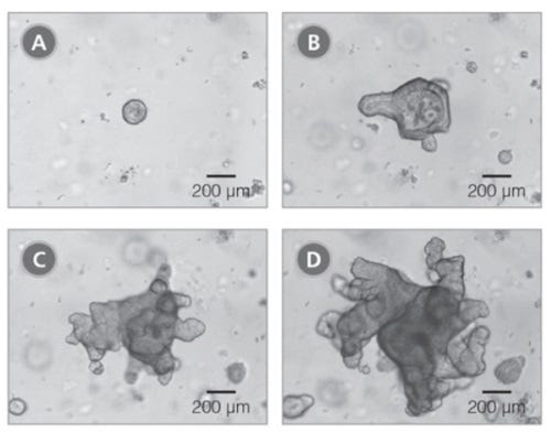
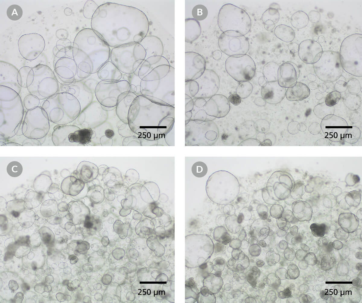
利用这种完整的培养基配方和优化方案建立和维持的肠道类器官来模拟成人肠上皮的关键特征。使用易于遵循和强大的方案,您可以在一周内从人类肠隐窝中获得类器官;在供体样本中,包括那些在其他情况下难以生长的样本,通过丰富的干细胞群可以实现类器官的生长。

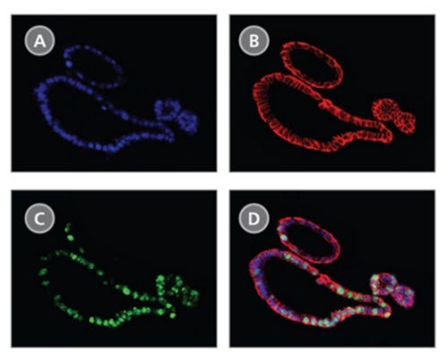
在intesteticult™类器官生长培养基(人类)中生长的类器官包含由极化肠上皮细胞层封闭的功能管腔,并且对于多功能建模应用,可以在3D或2D中进一步分化为浸入式单层或气液界面(ALI)培养interticult™类器官分化培养基(人类;目录# 100 - 0214)。

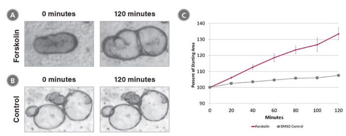
肠道类器官培养的应用包括研究肠上皮的发育和功能,建立肠道疾病模型,筛选肠道模型中的药效和毒性分子。肠道类器官培养也可用于研究成体干细胞特性和再生治疗方法。

学习如何培养人类肠道类器官按需肠道疗程或浏览我们的常见问题(FAQs)关于使用inteticult™的类器官工作流程。此外,下载我们详细的电子书经过验证的肠道类器官培养方案:开始使用inteticult™肠道类器官方案的精选集。

如果您打算将本产品用于商业用途,请与HUB Organoids B.V.联系www.huborganoids.nl申请商业用途许可证或澄清与HUB Organoids B.V.许可有关的信息。

Subtype
Specialized Media
 
Cell Type
Intestinal Cells
 
Species
Human
 
Application
Cell Culture, Differentiation, Expansion, Maintenance, Organoid Culture
 
Brand
IntestiCult
 
Area of Interest
Disease Modeling, Drug Discovery and Toxicity Testing, Epithelial Cell Biology, Stem Cell Biology
 

Data Figures

Figure 1. Primary Organoids Grown in IntestiCult™ Organoid Growth Medium (Human) are Fully Mature After 10-14 Days in Culture

Primary organoids were cultured from human colonic biopsy samples and grown in IntestiCult Organoid Growth Medium (Human). Organoids were imaged after (A) two days, (B) six days, (C) eight days and (D) ten days growth.

Figure 2. Organoids Grown in IntestiCult™ Organoid Growth Medium (Human) Display Markers of Human Intestinal Epithelial Cells

Immunofluorescence of organoids grown in IntestiCult™ Organoid Growth Medium (Human) showing colocalization of (A) DAPI, (B) EPCAM and (C) Ki67. (D) A merged image shows the position of actively proliferating (Ki67+) intestinal stem cells within the epithelial layer (EPCAM+).

Figure 3. Forskolin-Induced Swelling of Organoids Grown in IntestiCult™ Organoid Growth Medium (Human)

Organoids were treated with (A) 5 μM Forskolin or (B) DMSO and organoid area was measured at 0 minutes and 120 minutes. (C)Forskolin-treated organoids increased in size 33.5 ± 3.8% compared to 7.5 ± 0.8% for DMSO-treated organoids.

Figure 4. IntestiCult™ Organoid Growth Medium (Human) Supports the Growth of Organoids in Multiple Extracellular Matrices

Intestinal organoid cultures were prepared in IntestiCult™ Organoid Growth Medium (Human) and plated in (A) Matrigel® Growth Factor Reduced Basement Membrane Matrix (Corning® catalog # 356231), (B) Geltrex® LDEV-Free Reduced Growth Factor Basement Membrane Matrix (Gibco™ catalog # A1413202), (C) Cultrex® Reduced Growth Factor Basement Membrane Extract, Type 1 (R&D Systems™ catalog # 3433-005-R1), and (D) Cultrex® Reduced Growth Factor Basement Membrane Extract, Type 2 (R&D Systems™ catalog # 3533-005-02). Organoid cultures are imaged at the end of passage 4. All four extracellular matrices supported robust growth of human intestinal organoids. Scale bars = 250 μm.

Data and images of the MIMETAS OrganoReady Colon Organoid Plate

Figure 5. The MIMETAS OrganoReady® Colon Organoid Platform Uses IntestiCult™ to Create an Advanced Physiologically Relevant Model for Gastrointestinal Toxicity Testing and Barrier Integrity

(A) The OrganoReady® plate highlighting the microfluidic compartments.

(B) Schematic of the OrganoReady® microfluidic compartments where columns 1, 2, and 3 house the medium, a collagen-1 matrix, and the colon organoid tubule, respectively.

(C) Immunofluorescence staining of the colon organoid tubule confirms an adult tissue phenotype with the presence of goblet cells (Muc2), enterocytes (Occludin), and stem cells (Sox9). The 3D-lumenized structure provides apical (Ezrin) and basolateral (Integrin-β4) access to the polarized epithelium. Additionally, the organoid tubules show polarized and modulatable activity of expression of P-glycoprotein (Pgp).

(D) The OrganoReady® Colon Organoid platform supports toxicity testing, as demonstrated by dose-dependent measurements of TEER, LDH, and ATP following exposure to Afatinib (n = 4, N = 2). After 72 hrs of exposure, a dose dependent decrease in TEER, cytotoxicity, and cell viability was observed. For more information, please visit mimetas.com/en/organoready-organoid/.

Protocols and Documentation

Find supporting information and directions for use in the Product Information Sheet or explore additional protocols below.

Document Type
Product Name
Catalog #
Lot #
Language
Catalog #
06010
Lot #
All
Language
English
Document Type
Safety Data Sheet 1
Catalog #
06010
Lot #
All
Language
English
Document Type
Safety Data Sheet 2
Catalog #
06010
Lot #
All
Language
English
Document Type
Safety Data Sheet 3
Catalog #
06010
Lot #
All
Language
English
Document Type
Safety Data Sheet 4
Catalog #
06010
Lot #
All
Language
English

Applications

This product is designed for use in the following research area(s) as part of the highlighted workflow stage(s). Explore these workflows to learn more about the other products we offer to support each research area.

Resources and Publications

Educational Materials (62)

Brochure

Publications (26)

In vivo development of immune tissue in human intestinal organoids transplanted into humanized mice. C. Bouffi et al. Nature biotechnology 2023 6

Abstract

Human intestinal organoids (HIOs) derived from pluripotent stem cells provide a valuable model for investigating human intestinal organogenesis and physiology, but they lack the immune components required to fully recapitulate the complexity of human intestinal biology and diseases. To address this issue and to begin to decipher human intestinal-immune crosstalk during development, we generated HIOs containing immune cells by transplanting HIOs under the kidney capsule of mice with a humanized immune system. We found that human immune cells temporally migrate to the mucosa and form cellular aggregates that resemble human intestinal lymphoid follicles. Moreover, after microbial exposure, epithelial microfold cells are increased in number, leading to immune cell activation determined by the secretion of IgA antibodies in the HIO lumen. This in vivo HIO system with human immune cells provides a framework for future studies on infection- or allergen-driven intestinal diseases.
Catechol-O-Methyltransferase Loss Drives Cell-Specific Nociceptive Signaling via the Enteric Catechol-O-Methyltransferase/microRNA-155/Tumor Necrosis Factor ? Axis Q. Zhou et al. Gastroenterology 2023 4

Abstract

BACKGROUND & AIMS The etiology of abdominal pain in postinfectious, diarrhea-predominant irritable bowel syndrome (PI-IBS-D) is unknown, and few treatment options exist. Catechol-O-methyltransferase (COMT), an enzyme that inactivates and degrades biologically active catecholamines, plays an important role in numerous physiologic processes, including modulation of pain perception. Our objective was to determine the mechanism(s) of how decreased colonic COMT in PI-IBS-D patients contributes to the chronic abdominal pain phenotype after enteric infections. METHODS Colon neurons, epithelial cells, and macrophages were procured with laser capture microdissection from PI-IBS-D patients to evaluate cell-specific colonic COMT, microRNA-155 (miR-155), and tumor necrosis factor (TNF) ? expression levels compared to recovered patients (infection cleared: did not develop PI-IBS-D) and control individuals. COMT-/-, colon-specific COMT-/-, and miR-155-/- mice and human colonoids were used to model phenotypic expression of COMT in PI-IBS-D patients and to investigate signaling pathways linking abdominal pain. Citrobacter rodentium and trinitrobenzene sulfonic acid animal models were used to model postinflammatory changes seen in PI-IBS-D patients. RESULTS Colonic COMT levels were significantly decreased and correlated with increased visual analog scale abdominal pain ratings in PI-IBS-D patients compared to recovered patients and control individuals. Colonic miR-155 and TNF-? were increased in PI-IBS-D patients with diminished colonic COMT. COMT-/- mice had significantly increased expression of miR-155 and TNF-? in both colon tissues and dorsal root ganglia. Introduction of cV1q antibody (anti-TNF-?) into mice reversed visceral hypersensitivity after C rodentium and trinitrobenzene sulfonic acid. CONCLUSIONS Decreased colonic COMT in PI-IBS-D patients drives abdominal pain phenotypes via the COMT/miR-155/TNF-? axis. These important findings will allow new treatment paradigms and more targeted and personalized medicine approaches for gastrointestinal disorders after enteric infections.
3D bioprinted colorectal cancer models based on hyaluronic acid and signalling glycans. F. Cadamuro et al. Carbohydrate polymers 2023 2

Abstract

In cancer microenvironment, aberrant glycosylation events of ECM proteins and cell surface receptors occur. We developed a protocol to generate 3D bioprinted models of colorectal cancer (CRC) crosslinking hyaluronic acid and gelatin functionalized with three signalling glycans characterized in CRC, 3'-Sialylgalactose, 6'-Sialylgalactose and 2'-Fucosylgalactose. The crosslinking, performed exploiting azide functionalized gelatin and hyaluronic acid and 4arm-PEG-dibenzocyclooctyne, resulted in biocompatible hydrogels that were 3D bioprinted with commercial CRC cells HT-29 and patient derived CRC tumoroids. The glycosylated hydrogels showed good 3D printability, biocompatibility and stability over the time. SEM and synchrotron radiation SAXS/WAXS analysis revealed the influence of glycosylation in the construct morphology, whereas MALDI-MS imaging showed that protein profiles of tumoroid cells vary with glycosylation, indicating that sialylation and fucosylation of ECM proteins induce diverse alterations to the proteome of the tumoroid and surrounding cells.

更多信息

更多信息
Species Human
Legal Statement: This product was developed under a license to intellectual property owned by Hubrecht Organoid Technology (HUB). This product is sold for research use only. Purchase of this product does not include the right to use it for drug screening aiming for commercial gain, equipment validation, biobanking, or for other commercial purposes. Purchasers wishing to use the product for purposes other than basic research use should contact HUB at www.huborganoids.nl to obtain a further license. Purchasers may apply for a License from HUB, which will not be unreasonably withheld by HUB. PRODUCTS ARE FOR RESEARCH USE ONLY AND NOT INTENDED FOR HUMAN OR ANIMAL DIAGNOSTIC OR THERAPEUTIC USES UNLESS OTHERWISE STATED. FOR ADDITIONAL INFORMATION ON QUALITY AT STEMCELL, REFER TO WWW.STEMCELL.COM/COMPLIANCE.
版权 © 2025 STEMCELL Technologies 技术有限公司。保留所有权利。