产品号 #06010_C
用于建立和维持人肠道类器官的培养基
Organoids have truly expanded the limits of what's possible for in vitro studies of the intestinal epithelium. By providing optimized culture media and robust, approachable protocols, we are making these technologies more accessible to researchers.

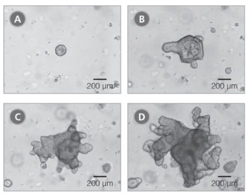
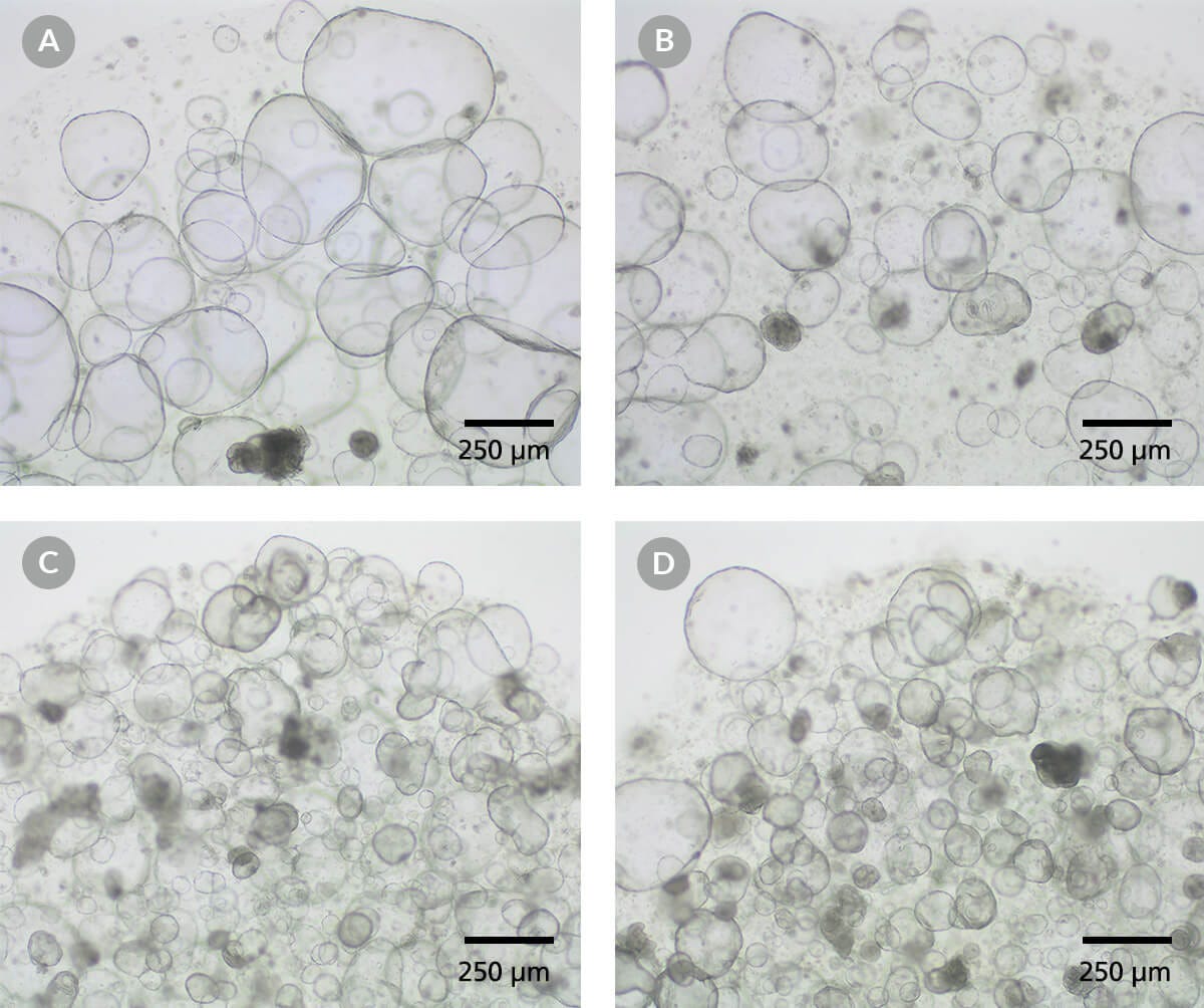
使用该完整培养基配方和优化的实验方案,构建和维持肠道类器官,以模拟成年肠上皮的关键特性。凭借该易于操作且有效的实验流程,可在一周内自人体肠隐窝衍生出类器官;富集的干细胞群促进了不同供体来源的类器官的生长,即使是原本较难培养的供体样本也能获得良好结果。
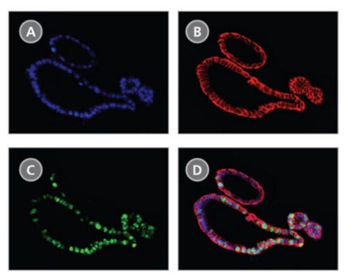
在IntestiCult™ 类器官生长培养基 (人)中生长的类器官包含一个由具有极性的肠上皮细胞层包围而成的功能性腔体结构。为满足多样化的建模应用,这些类器官可在三维结构中,或在浸没式单层培养或气液界面(ALI)培养的二维结构中,通过 IntestiCult™ 类器官分化培养基(人)(产品号 #100-0214)实现进一步分化。
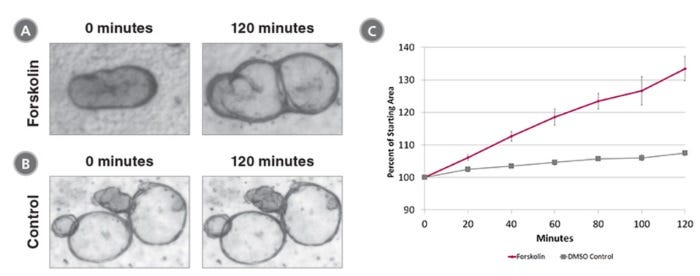
肠类器官培养的应用包括研究肠道上皮的发育和功能、构建肠道疾病模型,以及在肠道模型中筛选分子的有效性和毒性。肠类器官培养还可用于研究成体干细胞特性及再生治疗方法。
欢迎通过我们的免费点播课程学习人肠类器官的培养方法,或浏览关于使用 IntestiCult™ 进行类器官培养流程的常见问题(FAQs)。此外,您还可以下载我们的电子书《Proven Protocols for Intestinal Organoid Culture: Getting Started with IntestiCult™》,获取肠类器官培养方案的精选合集。
如果您打算将本产品用于商业目的,请通过www.huborganoids.nl与HUB Organoids B.V.联系,以获取商业用途许可或HUB Organoids B.V.许可的相关说明。
亚型
专用培养基
细胞类型
肠道细胞
种属
人
应用
细胞培养,分化,扩增,培养,类器官培养
品牌
IntestiCult
研究领域
疾病建模,药物发现和毒理检测,上皮细胞研究,干细胞生物学
Find supporting information and directions for use in the Product Information Sheet or explore additional protocols below.
This product is designed for use in the following research area(s) as part of the highlighted workflow stage(s). Explore these workflows to learn more about the other products we offer to support each research area.
| Species | Human |
|---|
用于将人肠道类器官在3D,或在单层/气液界面培养的形式下进一步分化的培养基
杜氏磷酸盐缓冲液,不含钙和镁离子
用于细胞分离和细胞培养的无菌聚丙烯锥形管