产品号 #100-0214_C
细胞培养基进一步分化人肠道类器官在三维,或作为单层/气液界面培养
Cell culture medium for establishment and maintenance of human intestinal organoids
cGMP, enzyme-free cell dissociation reagent
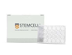
Polystyrene plate with lid and polyester membrane inserts for cell culture that feed basolaterally
RHO/ROCK pathway inhibitor; Inhibits ROCK1 and ROCK2
Organoids have truly expanded the limits of what's possible for in vitro studies of the intestinal epithelium. By providing optimized culture media and robust, approachable protocols, we are making these technologies more accessible to researchers.

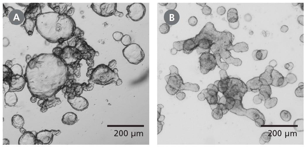
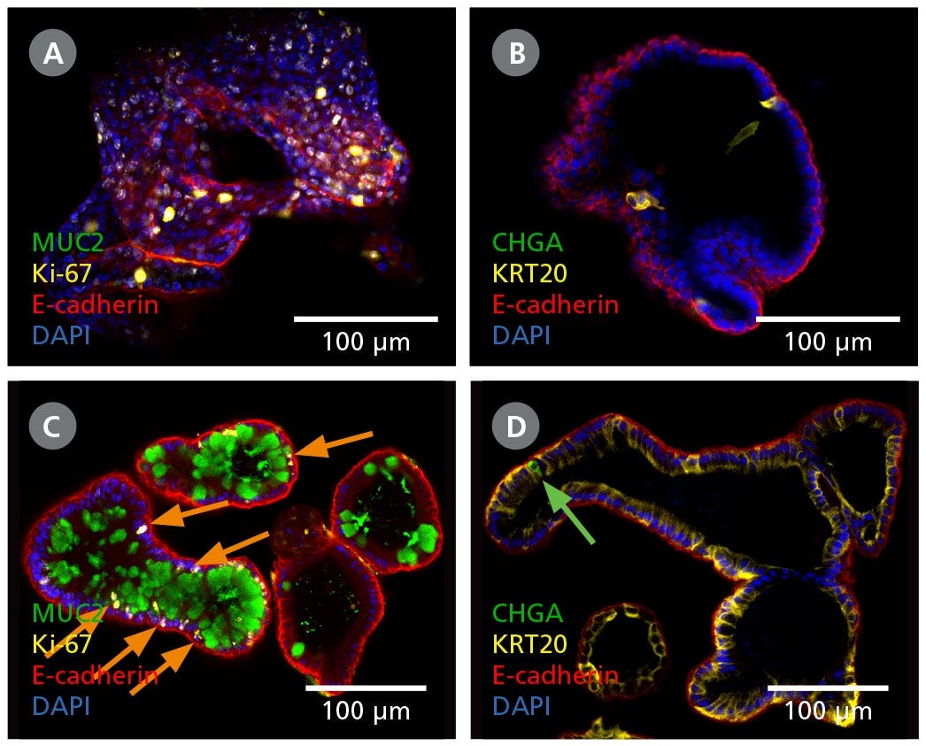
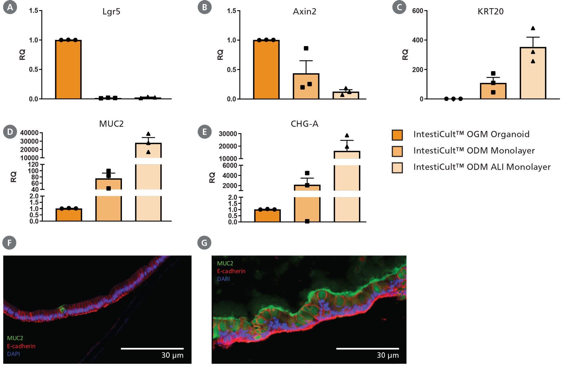
inteticult™类器官分化培养基(人)是一种完整的培养基,支持在三维(3D)或二维(单层或气液界面(ALI)培养中进一步分化肠道类器官。起始培养物可以是来源于人肠隐窝的类肠道器官,也可以是与interticult™类器官生长培养基(人类;目录# 06010)。
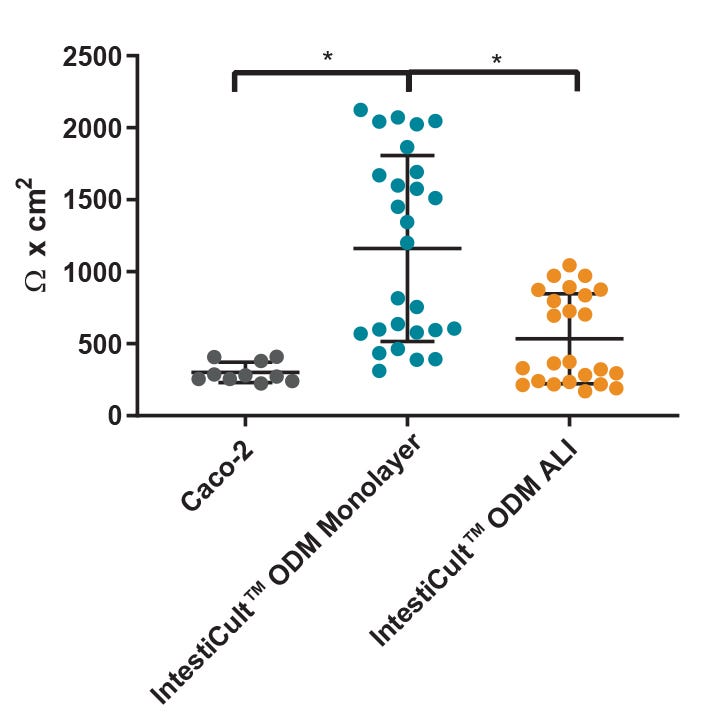
使用肠系膜™类器官分化培养基(人)生成的肠道培养物含有生理相关比例的分化细胞和干细胞群,概括了隐窝绒毛轴的多样性。与传统细胞系相比,肠单层细胞系表现出更大的屏障完整性,表达更高水平的关键分化标志物,并且具有更能代表体内肠道的形态学。
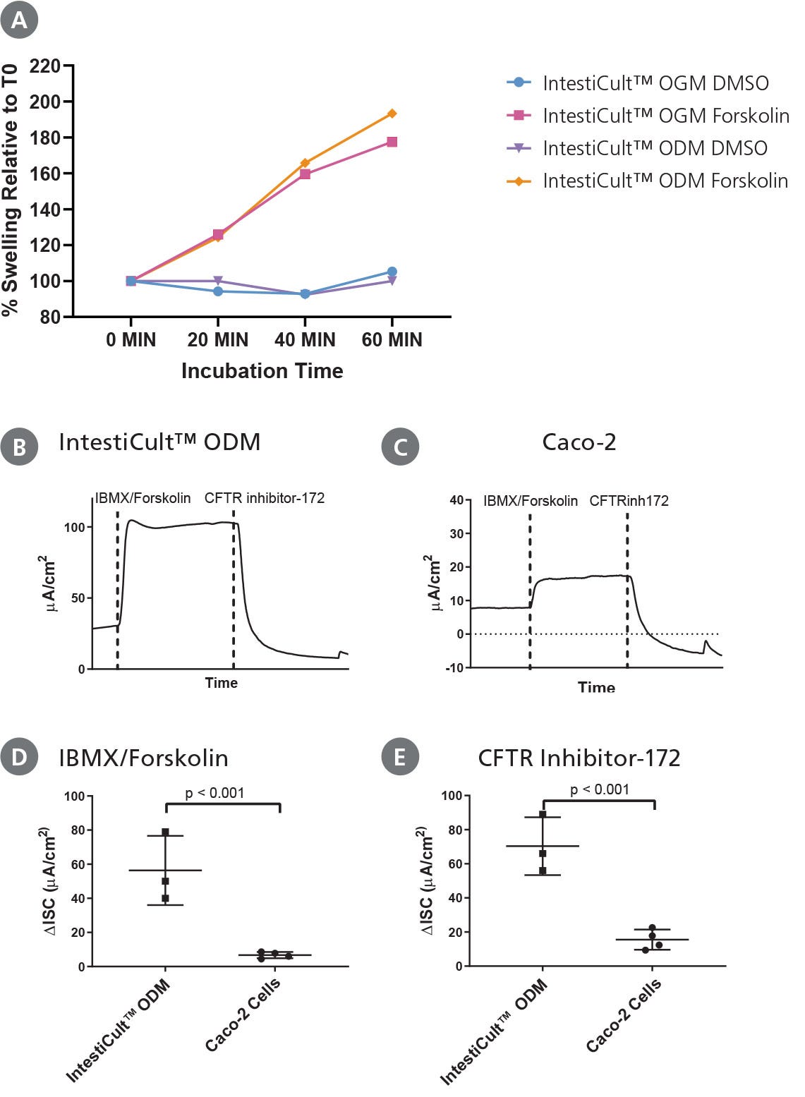
肠道类器官培养的应用包括研究肠上皮的发育和功能,模拟肠道疾病,化合物筛选和再生治疗方法。肠道单层培养物和ALI培养物特别适合于渗透性测定和传染病研究,因为它们易于接触到根尖表面。该试剂盒需要inteticult™类器官生长培养基(人;目录#06010),用于肠道类器官分化前的起始和扩张。
学习如何培养人类肠道类器官按需肠道疗程或浏览我们的常见问题(FAQs)关于使用inteticult™的类器官工作流程。此外,下载我们详细的电子书经过验证的肠道类器官培养方案:开始使用inteticult™肠道类器官方案的精选集。
如果您打算将本产品用于商业用途,请与HUB Organoids B.V.联系www.huborganoids.nl申请商业用途许可证或澄清与HUB Organoids B.V.许可有关的信息。
Subtype
Specialized Media
Cell Type
Intestinal Cells
Species
Human
Application
Cell Culture, Differentiation, Expansion, Maintenance, Organoid Culture
Brand
IntestiCult
Area of Interest
Disease Modeling, Drug Discovery and Toxicity Testing, Epithelial Cell Biology, Stem Cell Biology
Find supporting information and directions for use in the Product Information Sheet or explore additional protocols below.
This product is designed for use in the following research area(s) as part of the highlighted workflow stage(s). Explore these workflows to learn more about the other products we offer to support each research area.
| Species | Human |
|---|
酶解细胞试剂
Dulbecco's Modified Eagle's Medium/营养型火腿混合物F-12 (DMEM/F-12)与15 mM HEPES缓冲液
Dulbecco的磷酸盐缓冲盐水,不含钙和镁
Notch 通路抑制剂;抑制 γ-分泌酶