EasySep™人CD3正选试剂盒II

人CD3+细胞的免疫磁珠正选试剂盒

产品号 #(选择产品)

产品号 #17851_C

人CD3+细胞(如T细胞)的免疫磁珠正选

产品优势

  • 快速、易于操作
  • 纯度高达99%
  • 无需分离柱

产品组分包括

  • EasySep™人CD3正选试剂盒II(产品号 #17851)
  • EasySep™人CD3正选抗体混合物II,1mL
  • EasySep™ Dextran RapidSpheres™ 50100 磁珠,1mL
  • EasySep™人CD3正选试剂盒II(产品号 #100-0692)
  • EasySep™人CD3正选抗体混合物II,1 X 10mL
  • EasySep™ Dextran RapidSpheres™ 50100 磁珠,2 X 1mL
  • asySep™人CD3正选试剂盒II(产品号 #17851RF)
  • EasySep™人CD3正选抗体混合物II,1mL
  • EasySep™ Dextran RapidSpheres™ 50100 磁珠,1mL
  • RoboSep™ 缓冲液(产品号 #20104)
  • RoboSep™过滤吸头(产品号#20125)
New look, same high quality and support! You may notice that your instrument or reagent packaging looks slightly different from images displayed on the website, or from previous orders. We are updating our look but rest assured, the products themselves and how you should use them have not changed. Learn more

总览

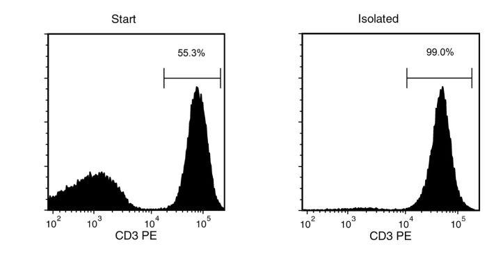
使用 EasySep™ 人 CD3 正选试剂盒 II,通过免疫磁珠正选,在短至 15 分钟内从新鲜或冻存的人外周血单核细胞 (PBMC) 或白细胞单采术样本中分离出高纯度的 CD3+ 细胞。EasySep™技术结合单克隆抗体的特异性和无需分离柱的简便磁分选系统,已在发表的研究中广泛应用超过20年。

在EasySep™正选过程中,目标细胞通过识别CD3的抗体复合物与磁珠进行标记。抗体复合物中还含有人Fc受体抗体,可最大程度地减少非特异性结合。使用EasySep™磁分选系统标记细胞,只需倾倒或吸出非目的细胞即可。目的细胞保留在管中。整个磁分选过程仅需15分钟,分选获得的CD3+细胞即可用于流式细胞术、细胞培养或DNA/RNA提取等下游应用。

该产品可替代EasySep™人CD3正选试剂盒 (产品号 #18051) 以实现更快更简单的细胞分选。

如需从白细胞分离样本中大规模分选人CD3+细胞,请选用大规格(1x10^10细胞)试剂盒(产品号#100-0692)。

了解更多关于免疫磁珠EasySep™技术的工作原理,或如何通过RoboSep™实现免疫磁珠细胞分选全自动化。探索为您的实验流程优化的更多产品,包括培养基、添加剂、抗体等。

磁体兼容性
• EasySep™ Magnet (Catalog #18000) • “The Big Easy” EasySep™ Magnet (Catalog #18001) • EasyPlate™ EasySep™ Magnet (Catalog #18102) • EasyEights™ EasySep™ Magnet (Catalog #18103) • Easy 50 EasySep™ Magnet (Catalog #18002) • RoboSep™-S (Catalog #21000) • Easy 250 EasySep™ Magnet (Catalog #100-0821)
 
亚型
细胞分选试剂盒
 
细胞类型
T 细胞
 
种属

 
样本来源
PBMC
 
筛选方法
Positive
 
应用
细胞分选
 
品牌
EasySep,RoboSep
 
研究领域
嵌合体,免疫,细胞治疗开发
 

Data Figures

Typical FACS Results with EasySep™ Human CD3 Positive Selection Kit II

Figure 1: Typical FACS Results with EasySep™ Human CD3 Positive Selection Kit II

Starting with a single cell suspension of human PBMCs, the CD3+ cell content of the isolated fraction is typically 99.2 ± 0.2% (mean ± SD), using the purple EasySep™ Magnet.

FACS Data for Anti-Human CD8a Antibody, Clone RPA-T8, FITC-Conjugated

Figure 2: FACS Data for Anti-Human CD8a Antibody, Clone RPA-T8, FITC-Conjugated

(A) Flow cytometry analysis of human peripheral blood mononuclear cells (PBMCs) labeled with Anti-Human CD8a Antibody, Clone RPA-T8, FITC (Catalog # 60022FI; filled histogram) or a mouse IgG1, kappa FITC isotype control antibody (solid line histogram). (B) Flow cytometry analysis of human PBMCs processed with the EasySep™ Human CD3 Positive Selection Kit (Catalog #17851) and labeled with Anti-Human CD8a Antibody, Clone RPA-T8, FITC. Histograms show labeling of PBMCs (Start) and isolated cells (Isolated). Labeling of start cells with a mouse IgG1, kappa FITC isotype control antibody is shown (solid line histogram).

FACS Data for Anti-Human CD4 Antibody, Clone OKT4, Alexa Fluor® 488-Conjugated

Figure 3: FACS Data for Anti-Human CD4 Antibody, Clone OKT4, Alexa Fluor® 488-Conjugated

(A) Flow cytometry analysis of human peripheral blood mononuclear cells (PBMCs) labeled with Anti-Human CD4 Antibody, Clone OKT4, Alexa Fluor® 488 (Catalog #60016AD) and Anti-Human CD45 Antibody, Clone HI30, APC (Catalog #60018AZ). (B) Flow cytometry analysis of human PBMCs isolated with the EasySep™ Human CD3 Positive Selection Kit (Catalog #17851) and labeled with Anti-Human CD4 Antibody, Clone OKT4, Alexa Fluor® 488. Histograms show labeling of total PBMCs (Start) and isolated cells (Isolated). Labeling with Mouse IgG2b, kappa Isotype Control Antibody, Clone MPC-11, Alexa Fluor® 488 (Catalog #60072AD) is shown in the bottom panel (solid line histogram). (C) Flow cytometry analysis of human PBMCs isolated with the EasySep™ Human CD4 Positive Selection Kit (Catalog #17852) and labeled with Anti-Human CD4 Antibody, Clone OKT4, Alexa Fluor® 488. Histograms show labeling of total PBMCs (Start) and isolated cells (Isolated). Labeling with Mouse IgG2b, kappa Isotype Control Antibody, Clone MPC-11, Alexa Fluor® 488 is shown in the bottom panel (solid line histogram).

Protocols and Documentation

Find supporting information and directions for use in the Product Information Sheet or explore additional protocols below.

Document Type
Product Name
Catalog #
Lot #
Language
Catalog #
17851RF
Lot #
All
Language
English
Catalog #
100-0692
Lot #
All
Language
English
Catalog #
17851
Lot #
All
Language
English
Catalog #
17851
Lot #
All
Language
English
Document Type
Safety Data Sheet 1
Catalog #
17851RF
Lot #
All
Language
English
Document Type
Safety Data Sheet 2
Catalog #
17851RF
Lot #
All
Language
English
Document Type
Safety Data Sheet 3
Catalog #
17851RF
Lot #
All
Language
English
Document Type
Safety Data Sheet 1
Catalog #
100-0692
Lot #
All
Language
English
Document Type
Safety Data Sheet 2
Catalog #
100-0692
Lot #
All
Language
English
Document Type
Safety Data Sheet 1
Catalog #
17851
Lot #
All
Language
English
Document Type
Safety Data Sheet 2
Catalog #
17851
Lot #
All
Language
English

Applications

This product is designed for use in the following research area(s) as part of the highlighted workflow stage(s). Explore these workflows to learn more about the other products we offer to support each research area.

Resources and Publications

Educational Materials (11)

Frequently Asked Questions

Can EasySep™ be used for either positive or negative selection?

Yes. The EasySep™ kits use either a negative selection approach by targeting and removing unwanted cells or a positive selection approach targeting desired cells. Depletion kits are also available for the removal of cells with a specific undesired marker (e.g. GlyA).

How does the separation work?

Magnetic particles are crosslinked to cells using Tetrameric Antibody Complexes (TAC). When placed in the EasySep™ Magnet, labeled cells migrate to the wall of the tube. The unlabeled cells are then poured off into a separate fraction.

Which columns do I use?

The EasySep™ procedure is column-free. That's right - no columns!

How can I analyze the purity of my enriched sample?

The Product Information Sheet provided with each EasySep™ kit contains detailed staining information.

Can EasySep™ separations be automated?

Yes. RoboSep™, the fully automated cell separator, automates all EasySep™ labeling and cell separation steps.

Can EasySep™ be used to isolate rare cells?

Yes. We recommend a cell concentration of 2x10⁸ cells/mL and a minimum working volume of 100 µL. Samples containing 2x10⁷ cells or fewer should be suspended in 100 µL of buffer.

Are the EasySep™ magnetic particles FACS-compatible?

Yes, the EasySep™ particles are flow cytometry-compatible, as they are very uniform in size and about 5000X smaller than other commercially available magnetic beads used with column-free systems.

Can the EasySep™ magnetic particles be removed after enrichment?

No, but due to the small size of these particles, they will not interfere with downstream applications.

Can I alter the separation time in the magnet?

Yes; however, this may impact the kit's performance. The provided EasySep™ protocols have already been optimized to balance purity, recovery and time spent on the isolation.

For positive selection, can I perform more than 3 separations to increase purity?

Yes, the purity of targeted cells will increase with additional rounds of separations; however, cell recovery will decrease.

How does the binding of the EasySep™ magnetic particle affect the cells? is the function of positively selected cells altered by the bound particles?

Hundreds of publications have used cells selected with EasySep™ positive selection kits for functional studies. Our in-house experiments also confirm that selected cells are not functionally altered by the EasySep™ magnetic particles.

If particle binding is a key concern, we offer two options for negative selection. The EasySep™ negative selection kits can isolate untouched cells with comparable purities, while RosetteSep™ can isolate untouched cells directly from whole blood without using particles or magnets.

Publications (4)

Immune responses against shared antigens are common in esophago-gastric cancer and can be enhanced using CD40-activated B cells. M. Thelen et al. Journal for immunotherapy of cancer 2022 dec

Abstract

BACKGROUND Specific immune response is a hallmark of cancer immunotherapy and shared tumor-associated antigens (TAAs) are important targets. Recent advances using combined cellular therapy against multiple TAAs renewed the interest in this class of antigens. Our study aims to determine the role of TAAs in esophago-gastric adenocarcinoma (EGA). METHODS RNA expression was assessed by NanoString in tumor samples of 41 treatment-na{\{i}}ve EGA patients. Endogenous T cell and antibody responses against the 10 most relevant TAAs were determined by FluoroSpot and protein-bound bead assays. Digital image analysis was used to evaluate the correlation of TAAs and T-cell abundance. T-cell receptor sequencing in vitro expansion with autologous CD40-activated B cells (CD40Bs) and in vitro cytotoxicity assays were applied to determine specific expansion clonality and cytotoxic activity of expanded T cells. RESULTS 68.3% of patients expressed ??5 TAAs simultaneously with coregulated clusters which were similar to data from The Cancer Genome Atlas (n=505). Endogenous cellular or humoral responses against ??1??TAA were detectable in 75.0% and 53.7% of patients respectively. We found a correlation of T-cell abundance and the expression of TAAs and genes related to antigen presentation. TAA-specific T-cell responses were polyclonal could be induced or enhanced using autologous CD40Bs and were cytotoxic in vitro. Despite the frequent expression of TAAs co-occurrence with immune responses was rare. CONCLUSIONS We identified the most relevant TAAs in EGA for monitoring of clinical trials and as therapeutic targets. Antigen-escape rather than missing immune response should be considered as mechanism underlying immunotherapy resistance of EGA."
Fibroblasts and macrophages cooperate to create a pro-tumorigenic and immune resistant environment via activation of TGF-$\beta$/IL-6 pathway in neuroblastoma. K. Louault et al. Oncoimmunology 2022

Abstract

Tumor-associated macrophages (TAM) and cancer-associated fibroblasts (CAF) and their precursor mesenchymal stromal cells (MSC) are often detected together in tumors, but how they cooperate is not well understood. Here, we show that TAM and CAF are the most abundant nonmalignant cells and are present together in untreated human neuroblastoma (NB) tumors that are also poorly infiltrated with T and natural killer (NK) cells. We then show that MSC and CAF-MSC harvested from NB tumors protected human monocytes (MN) from spontaneous apoptosis in an interleukin (IL)-6 dependent mechanism. The interactions of MN and MSC with NB cells resulted in a significant induction or increase in the expression of several pro-tumorigenic cytokines/chemokines (TGF-$\beta$1, MCP-1, IL-6, IL-8, and IL-4) but not of anti-tumorigenic cytokines (TNF-$\alpha$, IL-12) by MN or MSC, while also inducing cytokine expression in quiescent NB cells. We then identified a TGF-$\beta$1/IL-6 pathway where TGF-$\beta$1 stimulated the expression of IL-6 in NB cells and MSC, promoting TAM survival. Evidence for the contribution of TAM and MSC to the activation of this pathway was then provided in xenotransplanted NB tumors and patients with primary tumors by demonstrating a direct correlation between the presence of CAF and p-SMAD2 and p-STAT3. The data highlight a new mechanism of interaction between TAM and CAF supporting their pro-tumorigenic function in cancer.
BiHC, a T-Cell-Engaging Bispecific Recombinant Antibody, Has Potent Cytotoxic Activity Against Her2 Tumor Cells. Xing J et al. Translational oncology 2017 OCT

Abstract

Among different cancer immunotherapy approaches, bispecific antibodies (BsAbs) are of great interest due to their ability to recruit immune cells to kill tumor cells directly. Various BsAbs against Her2 tumor cells have been proposed with potent cytotoxic activities. However, most of these formats require extensive processing to obtain heterodimeric bispecific antibodies. In this study, we describe a bispecific antibody, BiHC (bispecific Her2-CD3 antibody), constructed with a single-domain anti-Her2 and a single-chain Fv (variable fragment) of anti-CD3 in an IgG-like format. In contrast to most IgG-like BsAbs, the two arms in BiHC have different molecular weights, making it easier to separate hetero- or homodimers. BiHC can be expressed in Escherichia coli and purified via Protein A affinity chromatography. The purified BiHC can recruit T cells and induce specific cytotoxicity of Her2-expressing tumor cells in vitro. The BiHC can also efficiently inhibit the tumor growth in vivo. Thus, BiHC is a promising candidate for the treatment of Her2-positive cancers.

更多信息

更多信息
Species Human
Magnet Compatibility • EasySep™ Magnet (Catalog #18000) • “The Big Easy” EasySep™ Magnet (Catalog #18001) • EasyPlate™ EasySep™ Magnet (Catalog #18102) • EasyEights™ EasySep™ Magnet (Catalog #18103) • Easy 50 EasySep™ Magnet (Catalog #18002) • RoboSep™-S (Catalog #21000) • Ea
Sample Source PBMC
Selection Method Positive
标记抗体
PRODUCTS ARE FOR RESEARCH USE ONLY AND NOT INTENDED FOR HUMAN OR ANIMAL DIAGNOSTIC OR THERAPEUTIC USES UNLESS OTHERWISE STATED. FOR ADDITIONAL INFORMATION ON QUALITY AT STEMCELL, REFER TO WWW.STEMCELL.COM/COMPLIANCE.
版权 © 2025 STEMCELL Technologies 技术有限公司。保留所有权利。