ImmunoCult™ -SF人巨噬细胞培养基

人单核细胞向巨噬细胞分化的无血清培养基

产品号 #(选择产品)

产品号 #10961_C

人单核细胞向巨噬细胞分化的无血清培养基

产品优势

  • 无血清培养基,无需额外补充血清;
  • 支持稳定的巨噬细胞分化;
  • 高产量得到巨噬细胞,具有理想的表型和功能;
  • 在6天后即可收集M1或M2巨噬细胞

总览

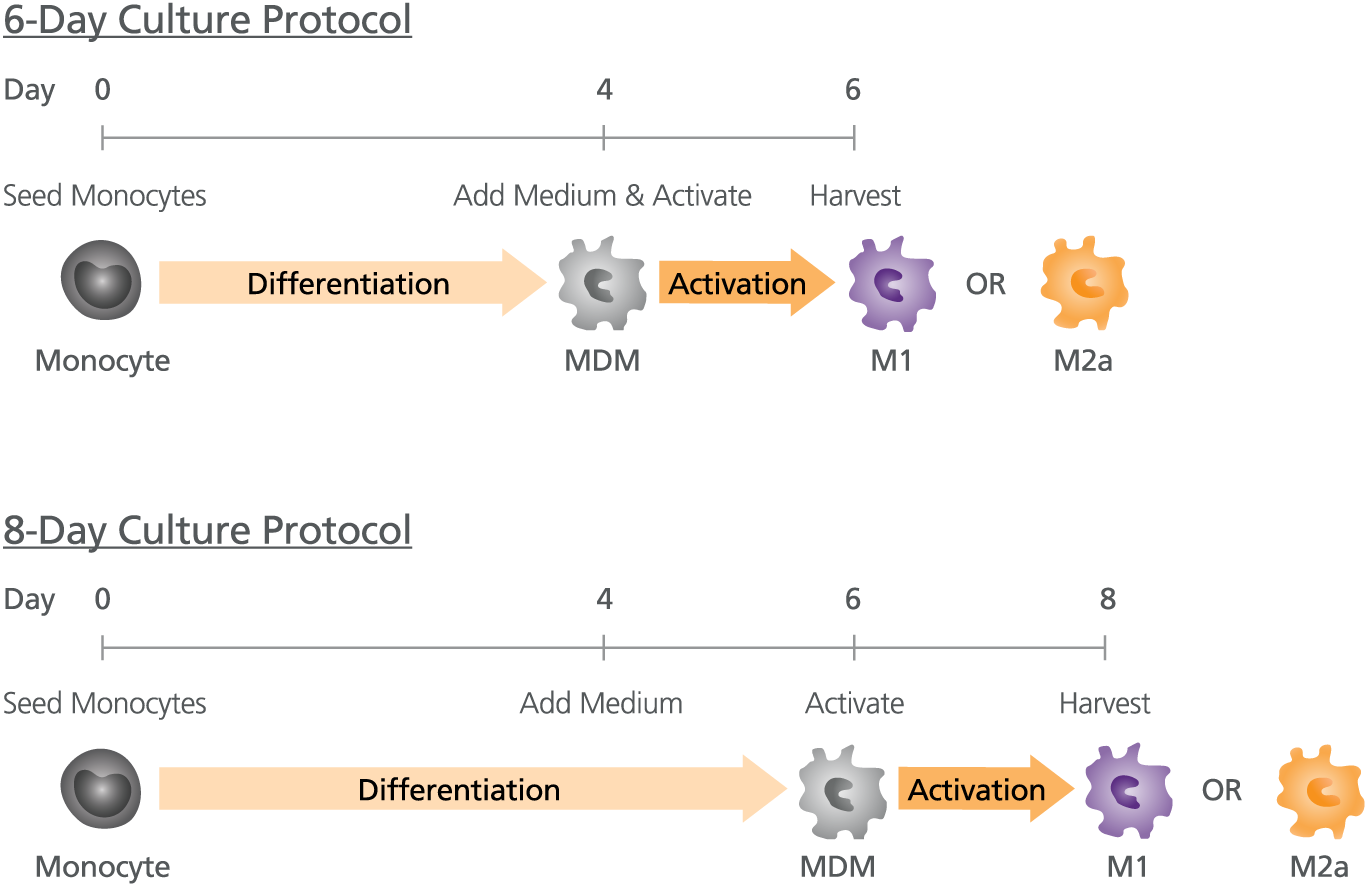
ImmunoCult™-SF巨噬细胞培养基用于搭配适当的细胞因子和刺激剂进行人单核细胞体外培养和向巨噬细胞分化。Immunocult™-SF巨噬细胞培养基中不包含巨噬细胞分化和活化因子,以便用户灵活地制备满足其要求的完全培养基。该培养基是一种专门的无血清培养基,可用于在6或8天周期的人单核细胞向M1(经典活化)和M2a(非传统活化)巨噬细胞分化。

包含
• Iscove’s MDM • Pre-tested bovine serum albumin • Recombinant human insulin • Human transferrin (iron-saturated) • 2-Mercaptoethanol • Supplements
 
亚型
专用培养基
 
细胞类型
巨噬细胞,单核细胞
 
种属

 
应用
细胞培养,分化
 
品牌
ImmunoCult
 
研究领域
免疫,感染性疾病(传染病)
 
制剂类别
无血清
 

Data Figures

Figure 1. Protocol for the Generation of M1 or M2a Activated Macrophages

Generate monocyte-derived macrophages (MDM) from isolated monocytes by culturing the cells in ImmunoCult™-SF Macrophage Differentiation Medium (ImmunoCult™-SF Macrophage Medium Catalog #10961 with added Human Recombinant M-CSF Catalog #78057). With our 8-day protocol, top-up with fresh ImmunoCult™-SF Macrophage Differentiation Medium on Day-4 and drive specific macrophage activation using appropriate stimuli on Day-6 (IFN-γ+LPS (Catalog #100-1270) for M1 activation and IL-4 for M2a activation). At Day-8 harvest fully mature M1 or M2a macrophages for use in downstream applications. With our 6-day protocol, macrophage activation can be done at the same time as the medium top-up step on Day-4 and harvested on Day-6.

Figure 2. ImmunoCult™-SF Supports Greater M1 and M2a Macrophage Yields Than Competitor’s Serum-Free Medium

Monocytes were cultured in ImmunoCult™-SF Macrophage Medium or a competitor’s serum-free macrophage medium and differentiated into macrophages using an 8-day protocol as shown in Figure 1. At Day-8, macrophages were harvested, counted and analysed by flow cytometry to assess the expression of macrophage markers CD80, CCR7, CD206 and CD209. (A) M1 macrophages were CD80+CCR7+ whereas (B) M2a macrophages showed a CD206+CD209+ phenotype. Macrophage yields are expressed as a percentage of total viable cells at Day 8 relative to the count of initial monocytes at Day 0. Macrophage yields were significantly higher in ImmunoCult™-SF than in Competitor’s serum-free medium (P < 0.05, paired t-test; mean ± SEM; n=18-19).

Figure 3. Activated Macrophages Generated with ImmunoCult™-SF Secrete the Appropriate Cytokines

Macrophages were generated with ImmunoCult™SF Macrophage Medium and activated using IFN-γ+LPS (Catalog #100-1270 ; M1) or IL-4 (M2a) in an 8-day protocol. At Day-8, supernatants from M1 and M2a macrophage cultures were collected and the concentrations of TNF-α, IL-12 (p70) and IL-10 were determined by ELISA. (A) M1 macrophages secreted 2821 ± 396 pg/ml TNF-α (n=24) and 656 ± 86 pg/mL IL-12 (p70) (n=25). (B) M2a macrophages produced 29 ± 6 pg/mL IL 10 (n=21) and did not produce TNF-α (below limit of detection, n=20). Data represents the mean ± SEM.

Protocols and Documentation

Find supporting information and directions for use in the Product Information Sheet or explore additional protocols below.

Document Type
Product Name
Catalog #
Lot #
Language
Catalog #
10961
Lot #
All
Language
English
Document Type
Safety Data Sheet
Catalog #
10961
Lot #
All
Language
English

Applications

This product is designed for use in the following research area(s) as part of the highlighted workflow stage(s). Explore these workflows to learn more about the other products we offer to support each research area.

Resources and Publications

Educational Materials (11)

Publications (1)

The Vi Capsular Polysaccharide of Salmonella Typhi Promotes Macrophage Phagocytosis by Binding the Human C-Type Lectin DC-SIGN. L. F. Zhang et al. mBio 2022 dec

Abstract

Capsular polysaccharides are common virulence factors of extracellular, but not intracellular bacterial pathogens, due to the antiphagocytic properties of these surface structures. It is therefore paradoxical that Salmonella enterica subspecies enterica serovar Typhi, an intracellular pathogen, synthesizes a virulence-associated (Vi) capsule, which exhibits antiphagocytic properties. Here, we show that the Vi capsular polysaccharide has different functions when S. Typhi interacts with distinct subsets of host phagocytes. The Vi capsular polysaccharide allowed S. Typhi to selectively evade phagocytosis by human neutrophils while promoting human macrophage phagocytosis. A screen of C-type lectin receptors identified human DC-SIGN as the receptor involved in macrophage binding and phagocytosis of capsulated S. Typhi. Consistent with the anti-inflammatory activity of DC-SIGN, purified Vi capsular polysaccharide reduced inflammatory responses in macrophages. These data suggest that binding of the human C-type lectin receptor DC-SIGN by the Vi capsular polysaccharide contributes to the pathogenesis of typhoid fever. IMPORTANCE Salmonella enterica subspecies enterica serovar Typhi is the causative agent of typhoid fever. The recent emergence of S. Typhi strains which are resistant to antibiotic therapy highlights the importance of vaccination in managing typhoid fever. The virulence-associated (Vi) capsular polysaccharide is an effective vaccine against typhoid fever, but the role the capsule plays during pathogenesis remains incompletely understood. Here, we identify the human C-type lectin receptor DC-SIGN as the receptor for the Vi capsular polysaccharide. Binding of capsulated S. Typhi to DC-SIGN resulted in phagocytosis of the pathogen by macrophages and induction of an anti-inflammatory cytokine response. Thus, the interaction of the Vi capsular polysaccharide with human DC-SIGN contributes to the pathogenesis of typhoid fever and should be further investigated in the context of vaccine development.

更多信息

更多信息
Species Human
Contains • Iscove’s MDM • Pre-tested bovine serum albumin • Recombinant human insulin • Human transferrin (iron-saturated) • 2-Mercaptoethanol • Supplements
Formulation Category Serum-Free
PRODUCTS ARE FOR RESEARCH USE ONLY AND NOT INTENDED FOR HUMAN OR ANIMAL DIAGNOSTIC OR THERAPEUTIC USES UNLESS OTHERWISE STATED. FOR ADDITIONAL INFORMATION ON QUALITY AT STEMCELL, REFER TO WWW.STEMCELL.COM/COMPLIANCE.
版权 © 2025 STEMCELL Technologies 技术有限公司。保留所有权利。