ImmunoCult™ 人NK扩增组套

人NK细胞培养扩增试剂盒

产品号 #(选择产品)

产品号 #100-0711_C

人NK细胞培养扩增试剂盒

产品优势

  • 高产量、高纯度的NK细胞稳定扩增;
  • 无饲料层细胞、无血清培养条件;
  • 扩增后的NK细胞具有细胞杀伤功能

产品组分包括

  • ImmunoCult™NK细胞基础培养基,500 mL(产品号#100-0712);
  • ImmunoCult™NK细胞扩增补充剂,5ml(产品号#100-0715);
  • ImmunoCult™NK细胞扩增包被材料,1.5 mL(产品号#100-0714)
专为您的实验方案打造的产品
要查看实验方案所需的所有配套产品,请参阅《实验方案与技术文档》
    string(7) "07050_C" NULL
  1. string(7) "37350_C" NULL
  2. string(7) "38040_C" NULL

总览

在无血清条件下持续扩增自然杀伤(NK)细胞,不使用可能带来后续问题的饲养层细胞。

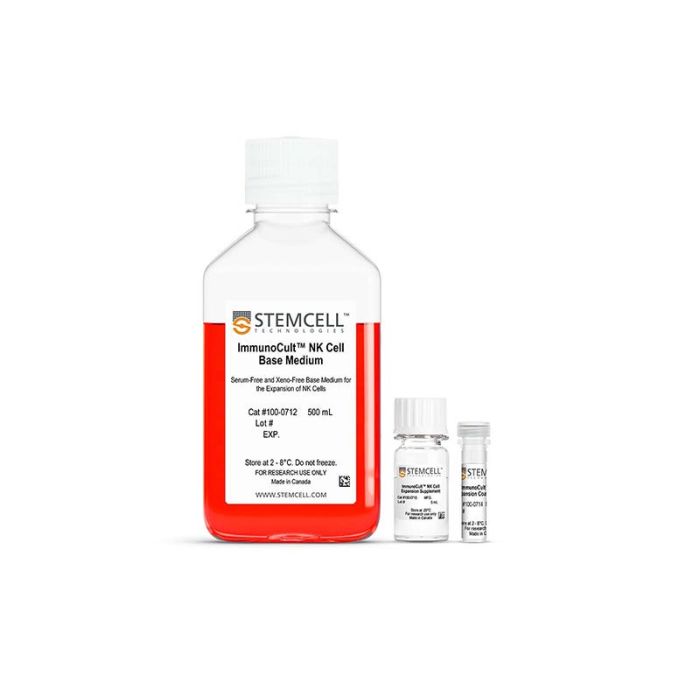
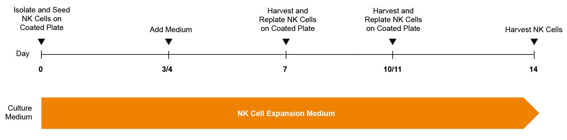
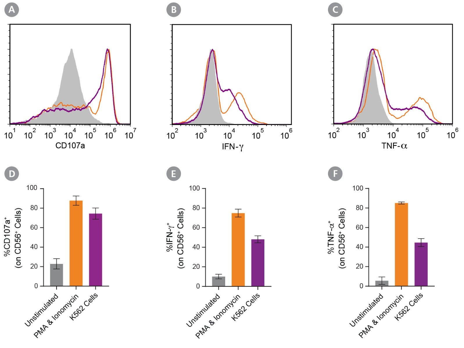
使用ImmunoCult™NK细胞扩增试剂盒,为高倍数扩增NK细胞提供优化的培养条件。该试剂盒包括ImmunoCult™NK细胞基础培养基,ImmunoCult™NK细胞扩增补充剂,和ImmunoCult™NK细胞扩增包被材料,为您提供完整且方便操作的培养体系。仅培养14天后,细胞就可以直接用于下游应用。

该试剂盒与我们的许多其他上下游产品兼容。例如,您可以使用EasySep™细胞分选试剂盒分选NK细胞,之后立即使用ImmunoCult™NK细胞扩增试剂盒扩增。

亚型
专用培养基
 
细胞类型
NK 细胞
 
种属

 
应用
细胞培养,扩增
 
品牌
ImmunoCult
 
研究领域
癌症,免疫,细胞治疗开发
 
制剂类别
Animal Component-Free,无血清,Xeno-Free
 

Data Figures

Protocol for the Expansion of Natural Killer (NK) Cells Using the ImmunoCult™ NK Cell Expansion Kit

Figure 1. ImmunoCult™ NK Cell Expansion Protocol

Human natural killer (NK) cells are isolated from blood or leukapheresis using EasySep™ selection. The NK cells are cultured in ImmunoCult™ NK Cell Expansion Medium, on plates coated with ImmunoCult™ NK Cell Expansion Coating Material. After 3 days, fresh medium is added to the culture. On day 7, and again on day 10 or 11, expanding NK cells are harvested and replated on freshly coated plates. Expanded NK cells were harvested on day 14 for use in downstream assays.

Cell Frequency, Fold Expansion, and Phenotyping of Natural Killer (NK) Cells Cultured Using the ImmunoCult™ NK Cell Expansion Kit

Figure 2. CD56+CD3− NK Cells Expand Over 14 Days in Feeder- and Serum-Free Culture Conditions

Isolated human CD56+CD3− NK cells were cultured using ImmunoCult™ NK Cell Expansion Kit for 14 days (Figure 1). Cells were harvested and analyzed for expression of characteristic NK cells markers, including CD56, CD3, CD16, CD94, KIR, NKG2D, NKp46, NKp30, and NKp44 by flow cytometry. Staining for killer cell immunoglobulin-like receptor (KIR) molecules was performed using two different antibody clones, HP-MA4 and 180704, which recognize distinct KIR molecules. Dead cells were excluded by light-scatter profile and DRAQ7™ staining. (A - H) Representative flow cytometry plots. (I) The average frequencies of viable CD56+CD3− and CD56+CD16+ NK cells on day 14 were 87 ± 1% and 75 ± 2%, respectively. The average fold expansion of CD56+CD3− cells was 89 ± 17. Results shown represent mean ± SEM (n = 34).

Cytotoxicity of Expanded Natural Killer (NK) Cells Co-Cultured with K562 Cells

Figure 3. Expanded NK Cells Are Functional, Killing K562 Cells in Co-Culture

Isolated CD56+CD3− NK cells were expanded as described in Figure 1. Expanded NK cells were co-cultured with Incucyte® Cytolight Rapid Dye-labeled K562 cells at 1:1 ratio of NK:K562 cells at 37°C for 4 hours. Incucyte® Caspase-3/7 Dye, a caspase-inducible dye, was added to the co-culture to detect caspase-induced apoptosis of the K562 cells. Images were obtained every hour using the Incucyte® imaging system and then analyzed to determine % killing (# apoptotic K562 cells ÷ # total labeled K562 cells). After 4 hours, an average of 48 ± 2.4% K562 cells were killed (n = 9). Data represent mean ± SEM.

Degranulation and Cytokine Production in Stimulated Natural Killer (NK) Cells Expanded Using the ImmunoCult™ NK Cell Expansion Kit

Figure 4. Expanded NK Cells Degranulate and Produce Cytokines After Stimulation

Isolated CD56+CD3− NK cells were expanded for 14 days (Figure 1). Expanded NK cells were left unstimulated (control) or were stimulated with either phorbol 12-myristate 13-acetate (PMA) and ionomycin or K562 cells at a ratio of 1:1 effector:target cells. CD107a antibody was added, and cultures were incubated at 37°C for 4 hours. After the first hour, Monensin and Brefeldin A were added. Cells were assessed for surface CD56, CD107a, and intracellular IFN-γ and TNF-α expression by flow cytometry. (A-C) Representative histograms of CD107a, IFN-γ, and TNF-α expression of unstimulated (grey filled), PMA and ionomycin-stimulated (orange), and K562-stimulated (purple) NK cell samples. (D) The average frequency of NK cells expressing surface CD107a, a marker of degranulation, was 23 ± 5% for the unstimulated control, 88 ± 5% after stimulation with PMA and ionomycin, and 74 ± 6% after stimulation with K562 cells. (E) The average frequency of NK cells expressing intracellular IFN-γ was 10 ± 2% for the unstimulated control, 75 ± 4% for cells stimulated with PMA and ionomycin, and 48 ± 4% for cells co-cultured with K562 cells. (F) The average frequency of NK cells expressing intracellular TNF-α was 6 ± 4% for the unstimulated control, 85 ± 1% cells stimulated with PMA and ionomycin, and 45 ± 4% for cells co-cultured with K562 cells. Data represent mean ± SEM (n = 6 - 13).

Protocols and Documentation

Find supporting information and directions for use in the Product Information Sheet or explore additional protocols below.

Document Type
Product Name
Catalog #
Lot #
Language
Catalog #
100-0711
Lot #
All
Language
English
Catalog #
100-0715
Lot #
All
Language
English
Catalog #
100-0712
Lot #
All
Language
English
Catalog #
100-0714
Lot #
All
Language
English
Document Type
Safety Data Sheet 1
Catalog #
100-0711
Lot #
All
Language
English
Document Type
Safety Data Sheet 2
Catalog #
100-0711
Lot #
All
Language
English
Document Type
Safety Data Sheet 3
Catalog #
100-0711
Lot #
All
Language
English
Document Type
Safety Data Sheet
Catalog #
100-0715
Lot #
All
Language
English
Document Type
Safety Data Sheet
Catalog #
100-0712
Lot #
All
Language
English
Document Type
Safety Data Sheet
Catalog #
100-0714
Lot #
All
Language
English

Applications

This product is designed for use in the following research area(s) as part of the highlighted workflow stage(s). Explore these workflows to learn more about the other products we offer to support each research area.

更多信息

更多信息
Species Human
Formulation Category Animal Component-Free, Serum-Free, Xeno-Free
For related products, including specialized culture and storage media, supplements, antibodies, cytokines, and small molecules, visit www.stemcell.com, or contact us at techsupport@stemcell.com.
版权 © 2025 STEMCELL Technologies 技术有限公司。保留所有权利。