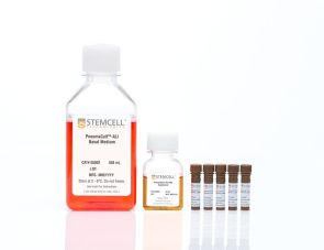
产品号 #100-1245_C
用于扩增和分化人输卵管类器官的无血清、支持性激素调节的模块化培养试剂盒
用于扩增和分化人输卵管类器官的无血清、支持性激素调节的模块化培养试剂盒
生成成熟的输卵管(fallopian tube, FT)类器官,呈现出与体内相似的纤毛细胞和分泌细胞的代表性组合。GyneCult™ 输卵管类器官培养基(FTOM)在功能性输卵管类器官培养中表现优于自配体系(DIY 配方),能够更好地支持相关细胞类型在生理比例下的生长。
由GyneCult™FTOM生成的类器官为女性生殖生物学研究提供了一个强大而通用的系统。通过该优化的工作流程,可以直接接种成3D培养从而节省时间和劳动力,并在第10天即可获得可传代的输卵管类器官。GyneCult™ FTOM 是一款无血清、性激素调节模块化的试剂盒;通过不添加 5000X 补充剂,用户可根据需要灵活调节激素水平,同时为特定应用提供一致、可重复的实验结果。
输卵管类器官培养的应用包括研究生育机制、激素动力学、月经及排卵后周期,以及卵巢癌。借助 GyneCult™ FTOM,可培养部分高等级浆液性卵巢癌(HGSOC)类器官,用于卵巢癌研究,例如评估输卵管上皮细胞的肿瘤发生能力,或用于卵巢癌化疗药物的筛选。
亚型
专用培养基
细胞类型
上皮细胞
应用
细胞培养,分化,扩增,类器官培养
品牌
GyneCult
研究领域
癌症,疾病建模,药物发现和毒理检测,上皮细胞研究,类器官
制剂类别
无血清

Figure 1. Generation of Fallopian Tube Organoids Using GyneCult™ Fallopian Tube Organoid Medium
(A) Workflow showing the generation of fallopian tube (FT) organoids. Freshly isolated or cryopreserved human FT cells are embedded at 4000 cells per 20 μL Matrigel® dome into 24-well non-tissue culture plates. Once the domes solidify, 500 μL of GyneCult™ Fallopian Tube Organoid Medium (FTOM) is added. By Day 10, cultures contain functional cells with beating cilia and can be passaged for further culture or cryopreservation. (B) Representative images of FT organoid morphologies throughout the GyneCult™ FTOM workflow. Scale bar = 200 μm.

Figure 2. GyneCult™ Fallopian Tube Organoid Medium Generates Consistent and Standardized Organoid Cultures
Dissociated human FT cells were seeded and cultured following the recommended protocol at 4000 cells per dome. Organoid cultures from passage 0 (A, D, G), passage 3 (B, E, H), and passage 5 (C, F, I), at 10 - 14 days post-seeding, are shown for three independent donors. Within each dome, FT organoids were typically cystic, with a prominent lumen and between 50 - 500 µm in diameter. Size distribution and diverse morphology within each culture were maintained from passage to passage. GyneCult™ FTOM supports human FT cultures with high consistency between donors and low passage-to-passage variability, while also preserving the morphological heterogeneity within each culture. Scale bar = 200 µm.

Figure 3. GyneCult™ Fallopian Tube Organoid Medium Generates Robust Cultures Without the Need for Additional Purification Steps
GyneCult™ FTOM supports passaging FT organoids as single cells or clumps and demonstrates a high establishment rate in the donors tested (n = 8), even with cultures that show poor initiation capabilities (e.g. Donor 3, 5 and 7). (A) FT organoids imaged at passage 3 (Donor 1), passage 4 (Donor 2), and passage 5 (Donors 3 - 8). Scale bar = 500 µm. (B) Representative flow cytometric dot plot of a dissociated donor sample stained with EpCAM and CD45 antibodies. Stromal or immune cell contamination could be present and is selected out with subsequent passages in GyneCult™ FTOM without additional purification steps. (C) Box plot showing the organoid-forming efficiency (OFE; the fraction of single cells that form organoids in culture) of dissociated FT cells is 16 ± 8% when normalized to the EpCAM+ population (mean ± SD, n = 10, includes 2 replicates).

Figure 4. GyneCult™ FTOM Supports Proliferation from Fresh or Cryopreserved Samples
GyneCult™ FTOM supports robust organoid formation from cryopreserved or freshly isolated FT cells. (A) Organoid morphology after establishing cultures from freshly dissociated single cells or revived after cryopreservation. Shown are organoids at the end of primary culture (top) and after passage 3 (bottom). Scale bar = 500 µm. (B) Throughout the 10 - 13 day primary passage, the proliferative capacity of fresh and cryopreserved cultures is expressed in doublings per day, with no significant difference. (C) Additionally, cultured FT cells retain their organoid forming efficiency (OFE) after thawing, fluctuating between 5 - 10%, as seen in the box and whiskers plot for at least five passages. (D) Graph showing individual cryopreserved FT donors (n = 7), the samples were able to undergo 20 cumulative doublings within 5 passages, further demonstrating the ability to achieve strong organoid cultures from cryopreserved samples.

Figure 5. Differentiated Fallopian Tube Organoids Exhibit a Representative Mixture of Ciliated and Secretory Cell Types
Dissociated primary FT cells from three individual donors were cultured in GyneCult™ FTOM and cultured for five passages. The resulting organoids were representative of in vivo FT biology, containing a mixture of ciliated and secretory cells as confirmed by marker antigen expression. (A-C) Cultures were stained for the secretory transcription factor PAX8 (green), and a functional component in ciliated cells—acetylated tubulin (red). (D-F) FT organoids were also stained for the secretory glycoprotein OVGP1 (green) and ciliated cell transcription factor FOXJ1 (red). These results demonstrate the efficient generation of multilineage organoids across different donors, mimicking the in vivo biology. Scale bar = 50 µm.

Figure 6. GyneCult™ FTOM Supports Robust Growth and High Fidelity Modeling Compared to DIY Medium
FT cells from the same donor were seeded and cultured side-by-side using (A) DIY medium and (D) GyneCult™ FTOM. The organoids were stained with IHC for (B, E) secretory transcription factor, PAX8, and (C, F) ciliated cell transcription factor, FOXJ1, both shown in brown. Blue stains indicate nuclei and the absence of nuclear expression of the target antigen. The DIY-derived organoids were dominated by (B) PAX8+ and (C) lacked FOXJ1+ markers, suggesting that these organoids consist mainly of secretory cells. (E, F) GyneCult™-derived organoids exhibited a representative mix of secretory and ciliated cells as it occurs in vivo. This demonstrates that GyneCult™ FTOM offers (D) superior growth and (E, F) multilineage representation when compared head-to-head with the DIY formulation. Data was used with permission from the Huntsman Lab, BC Cancer Research Center.

Figure 7. A Subset of High-Grade Serous Ovarian Cancer Organoids Can Be Cultured in GyneCult™ FTOM and Recapitulate Tumor Biology in 3D
(A) Representative images of a cryopreserved high-grade serous ovarian cancer (HGSOC) sample cultured in GyneCult™ FTOM, without the addition of the 100x supplement. (B) The resulting HGSOC-derived organoids exhibited a distinct morphology from that of healthy fallopian tube organoids, and (C) expressed HGSOC markers, such as PAX8 (green). (D) Throughout their culture, the HGSOC samples displayed robust growth and an increase in cumulative doublings within five passages. GyneCult™ FTOM supports the growth of a subset of HGSOC samples as organoid cultures to model tumor biology.
Find supporting information and directions for use in the Product Information Sheet or explore additional protocols below.
This product is designed for use in the following research area(s) as part of the highlighted workflow stage(s). Explore these workflows to learn more about the other products we offer to support each research area.
Thank you for your interest in IntestiCult™ Organoid Growth Medium (Human). Please provide us with your contact information and your local representative will contact you with a customized quote. Where appropriate, they can also assist you with a(n):
Estimated delivery time for your area
Product sample or exclusive offer
In-lab demonstration
| Formulation Category | Serum-Free |
|---|

在气液界面培养的人气道上皮细胞的无血清和无bpe培养基

用于 CFU 检测人乳腺上皮细胞的培养

用于培养人、小鼠和大鼠上皮干细胞
扫描二维码或搜索微信号STEMCELLTech,即可关注我们的微信平台,第一时间接收丰富的技术资源和最新的活动信息。
如您有任何问题,欢迎发消息给STEMCELLTech微信公众平台,或与我们通过电话/邮件联系:400 885 9050 INFO.CN@STEMCELL.COM。

