产品号 #85415_C
用于体外诊断(IVD)应用的密度梯度离心管
用于体外诊断(IVD)应用的密度梯度离心管
Density gradient medium for the isolation of mononuclear cells
Cell culture buffer
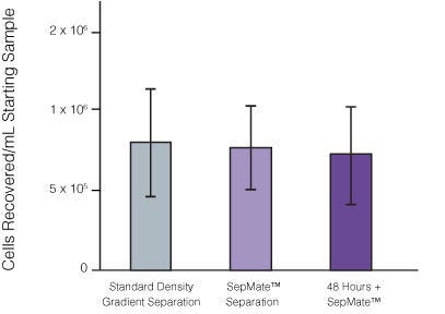
Traditional isolation of PBMCs requires careful layering of blood onto density gradient media prior to centrifugation. We developed SepMate™ to simplify this process, so anyone can isolate PBMCs with a simple pour while maintaining consistency across samples.

通过将SepMate™纳入您的密度梯度离心步骤,简化外周血单个核细胞(PBMC)分离。
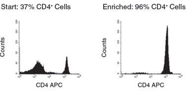
SepMate™管包含一个插入物,在密度梯度介质和血液之间形成屏障,从而消除了仔细分层血液的需要,并允许简单的倒液即可轻松收获单个核细胞。本品可与RosetteSep™分离特异性免疫细胞亚群。
SepMate™在cGMP下生产,并在澳大利亚、加拿大、欧洲和美国注册为体外诊断(IVD)设备。在中国,SepMate™被中国食品药品监督管理局(CFDA)视为非医疗器械,应作为一般实验室设备使用。最终用户负责确定产品是否适合他们的特定应用。
浏览我们的常见问题(FAQs)在SepMate™。
Contains
Find supporting information and directions for use in the Product Information Sheet or explore additional protocols below.
This product is designed for use in the following research area(s) as part of the highlighted workflow stage(s). Explore these workflows to learn more about the other products we offer to support each research area.
| Species | Human |
|---|---|
| Contains | Polypropylene tube containing an insert |
| Sample Source | Bone Marrow, Whole Blood |
| Selection Method | Negative |
免疫密度阴性选择鸡尾酒