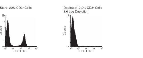
RosetteSep™人CD3去除抗体混合物

免疫密度去除试剂混合物

产品号 #(选择产品)

产品号 #15621_C

免疫密度梯度离心法去除CD3+T细胞

产品优势

  • 快捷、操作简单
  • 不需要特殊设备或额外培训
  • 获得的活细胞无标记
  • 可与SepMate™联合使用,实现一致的高通量样本处理

产品组分包括

  • RosetteSep™人CD3去除抗体混合物(产品号 #15624)
    • RosetteSep™人CD3去除抗体混合物,2mL
  • RosetteSep™人CD3去除抗体混合物(产品号 #15624)
    • RosetteSep™人CD3去除抗体混合物,5x2mL
专为您的实验方案打造的产品
要查看实验方案所需的所有配套产品,请参阅《实验方案与技术文档》

总览

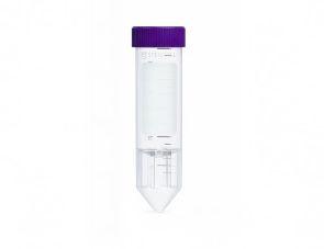
RosetteSep™人CD3去除混合物从全血中去除CD3+细胞。四聚体抗体复合物可识别CD3红细胞(RBC)上的糖蛋白A,从而靶向去除非目的细胞。使用密度梯度离心液如RosetteSep™ DM-L(产品号 #15705)或Lymphoprep™(产品号 #07801)离心后,非目的细胞会与红细胞一起沉淀。去除了CD3+T细胞的目的细胞为血浆和密度梯度离心液的交界界面中高度富集的细胞。

亚型
细胞分选试剂盒
 
细胞类型
T 细胞
 
种属

 
样本来源
Buffy Coat,Whole Blood
 
筛选方法
删除
 
应用
细胞分选
 
品牌
RosetteSep
 
研究领域
免疫
 

Data Figures

FACS Histogram Results Using RosetteSep™ Human CD3+ Cell Depletion Cocktail

Figure 1. FACS Histogram Results Using RosetteSep™ Human CD3+ Cell Depletion Cocktail

Protocols and Documentation

Find supporting information and directions for use in the Product Information Sheet or explore additional protocols below.

Document Type
Product Name
Catalog #
Lot #
Language
Catalog #
15621, 15661
Lot #
All
Language
English
Document Type
Safety Data Sheet
Catalog #
15621, 15661
Lot #
All
Language
English

Applications

This product is designed for use in the following research area(s) as part of the highlighted workflow stage(s). Explore these workflows to learn more about the other products we offer to support each research area.

Resources and Publications

Educational Materials (4)

Frequently Asked Questions

What is RosetteSep™?

RosetteSep™ is a rapid cell separation procedure for the isolation of purified cells directly from whole blood, without columns or magnets.

How does RosetteSep™ work?

The antibody cocktail crosslinks unwanted cells to red blood cells (RBCs), forming rosettes. The unwanted cells then pellet with the free RBCs when centrifuged over a density centrifugation medium (e.g. Ficoll-Paque™ PLUS, Lymphoprep™).

What factors affect cell recovery?

The temperature of the reagents can affect cell recovery. All reagents should be at room temperature (sample, density centrifugation medium, PBS, centrifuge) before performing the isolations. Layering can also affect recovery so be sure to carefully layer the sample to avoid mixing with the density centrifugation medium as much as possible. Be sure to collect the entire enriched culture without disturbing the RBC pellet. A small amount of density centrifugation medium can be collected without worry.

Which cell samples can RosetteSep™ be used with?

RosetteSep™ can be used with leukapheresis samples, bone marrow or buffy coat, as long as: the concentration of cells does not exceed 5 x 10⁷ per mL (can dilute if necessary); and there are at least 100 RBCs for every nucleated cell (RBCs can be added if necessary).

Can RosetteSep™ be used with previously frozen or cultured cells?

Yes. Cells should be re-suspended at 2 - 5 x 10⁷ cells / mL in PBS + 2% FBS. Fresh whole blood should be added at 250 µL per mL of sample, as a source of red cells.

Can RosetteSep™ be used to enrich progenitors from cord blood?

Yes. Sometimes cord blood contains immature nucleated red cells that have a lower density than mature RBCs. These immature red cells do not pellet over Ficoll™, which can lead to a higher RBC contamination than peripheral blood separations.

Does RosetteSep™ work with mouse cells?

No, but we have developed EasySep™, a magnetic-based cell isolation system which works with mouse and other non-human species.

Which anticoagulant should be used with RosetteSep™?

Peripheral blood should be collected in heparinized Vacutainers. Cord blood should be collected in ACD.

Should the anticoagulant be washed off before using RosetteSep™?

No, the antibody cocktail can be added directly to the sample.

Publications (8)

IFN-I and IL-22 mediate protective effects of intestinal viral infection. J. A. Neil et al. Nature microbiology 2019

Abstract

Products derived from bacterial members of the gut microbiota evoke immune signalling pathways of the host that promote immunity and barrier function in the intestine. How immune reactions to enteric viruses support intestinal homeostasis is unknown. We recently demonstrated that infection by murine norovirus (MNV) reverses intestinal abnormalities following depletion of bacteria, indicating that an intestinal animal virus can provide cues to the host that are typically attributed to the microbiota. Here, we elucidate mechanisms by which MNV evokes protective responses from the host. We identify an important role for the viral protein NS1/2 in establishing local replication and a type I interferon (IFN-I) response in the colon. We further show that IFN-I acts on intestinal epithelial cells to increase the proportion of CCR2-dependent macrophages and interleukin (IL)-22-producing innate lymphoid cells, which in turn promote pSTAT3 signalling in intestinal epithelial cells and protection from intestinal injury. In addition, we demonstrate that MNV provides a striking IL-22-dependent protection against early-life lethal infection by Citrobacter rodentium. These findings demonstrate novel ways in which a viral member of the microbiota fortifies the intestinal barrier during chemical injury and infectious challenges.
Comparative transcriptomic profile of tolerogenic dendritic cells differentiated with vitamin D3, dexamethasone and rapamycin. J. Navarro-Barriuso et al. Scientific reports 2018 OCT

Abstract

Tolerogenic dendritic cell (tolDC)-based therapies have become a promising approach for the treatment of autoimmune diseases by their potential ability to restore immune tolerance in an antigen-specific manner. However, the broad variety of protocols used to generate tolDC in vitro and their functional and phenotypical heterogeneity are evidencing the need to find robust biomarkers as a key point towards their translation into the clinic, as well as better understanding the mechanisms involved in the induction of immune tolerance. With that aim, in this study we have compared the transcriptomic profile of tolDC induced with either vitamin D3 (vitD3-tolDC), dexamethasone (dexa-tolDC) or rapamycin (rapa-tolDC) through a microarray analysis in 5 healthy donors. The results evidenced that common differentially expressed genes could not be found for the three different tolDC protocols. However, individually, CYP24A1, MUCL1 and MAP7 for vitD3-tolDC; CD163, CCL18, C1QB and C1QC for dexa-tolDC; and CNGA1 and CYP7B1 for rapa-tolDC, constituted good candidate biomarkers for each respective cellular product. In addition, a further gene set enrichment analysis of the data revealed that dexa-tolDC and vitD3-tolDC share several immune regulatory and anti-inflammatory pathways, while rapa-tolDC seem to be playing a totally different role towards tolerance induction through a strong immunosuppression of their cellular processes.
Ethyl Pyruvate Stimulates Regulatory T Cells and Ameliorates Type 1 Diabetes Development in Mice. I. Koprivica et al. Frontiers in immunology 2018

Abstract

Type 1 diabetes (T1D) is an autoimmune disease in which a strong inflammatory response causes the death of insulin-producing pancreatic beta-cells, while inefficient regulatory mechanisms allow that response to become chronic. Ethyl pyruvate (EP), a stable pyruvate derivate and certified inhibitor of an alarmin-high mobility group box 1 (HMGB1), exerts anti-oxidant and anti-inflammatory properties in animal models of rheumatoid arthritis and encephalomyelitis. To test its therapeutic potential in T1D, EP was administered intraperitoneally to C57BL/6 mice with multiple low-dose streptozotocin (MLDS)-induced T1D. EP treatment decreased T1D incidence, reduced the infiltration of cells into the pancreatic islets and preserved beta-cell function. Apart from reducing HMGB1 expression, EP treatment successfully interfered with the inflammatory response within the local pancreatic lymph nodes and in the pancreas. Its effect was restricted to boosting the regulatory arm of the immune response through up-regulation of tolerogenic dendritic cells (CD11c+CD11b-CD103+) within the pancreatic infiltrates and through the enhancement of regulatory T cell (Treg) levels (CD4+CD25highFoxP3+). These EP-stimulated Treg displayed enhanced suppressive capacity reflected in increased levels of CTLA-4, secreted TGF-beta, and IL-10 and in the more efficient inhibition of effector T cell proliferation compared to Treg from diabetic animals. Higher levels of Treg were a result of increased differentiation and proliferation (Ki67+ cells), but also of the heightened potency for migration due to increased expression of adhesion molecules (CD11a and CD62L) and CXCR3 chemokine receptor. Treg isolated from EP-treated mice had the activated phenotype and T-bet expression more frequently, suggesting that they readily suppressed IFN-gamma-producing cells. The effect of EP on Treg was also reproduced in vitro. Overall, our results show that EP treatment reduced T1D incidence in C57BL/6 mice predominantly by enhancing Treg differentiation, proliferation, their suppressive capacity, and recruitment into the pancreas.

更多信息

更多信息
Species Human
Sample Source Buffy Coat, Whole Blood
Selection Method Depletion
PRODUCTS ARE FOR RESEARCH USE ONLY AND NOT INTENDED FOR HUMAN OR ANIMAL DIAGNOSTIC OR THERAPEUTIC USES UNLESS OTHERWISE STATED. FOR ADDITIONAL INFORMATION ON QUALITY AT STEMCELL, REFER TO WWW.STEMCELL.COM/COMPLIANCE.
版权 © 2025 STEMCELL Technologies 技术有限公司。保留所有权利。