ReLeSR™ passaging protocol eliminates difficult and time-consuming steps, thereby enabling easy culture scale-up. Surface area of 4 x 6 well plates (230 cm²) is comparable to that of a T225 flask (225 cm²). TeSR™ = TeSR™ family media (mTeSR™1, TeSR™2, or TeSR™-E8™).
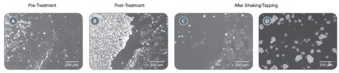
ReLeSR™ selectively detaches undifferentiated cells from pluripotent stem cell cultures without manual selection. Optimally-sized aggregates are generated following shaking/tapping of the cultureware. (A) An hPSC culture ready for passaging. Note the presence of differentiated cells at the edge of the undifferentiated hPSC colony. (B) Following incubation with ReLeSR™, the undifferentiated hPSC colony starts to lift off of the cultureware. The differentiated cells remain attached to the cultureware. (C) Following shaking/tapping of the cultureware, the undifferentiated cells completely lift off of the cultureware. (D) The undifferentiated hPSC colony is broken up into optimally-sized aggregates for replating.
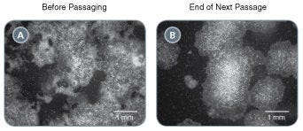
Figure 3. Rescue Highly Differentiated Cultures
Poor quality human pluripotent stem cell cultures containing large proportions of differentiated cells can be rescued by passaging with ReLeSR™. (A) A poor quality hPSC culture containing ~50% undifferentiated cells. (B) Following passaging with ReLeSR™, the differentiated cells have largely been eliminated from the culture, with >90% undifferentiated cells present at the end of the next passage.
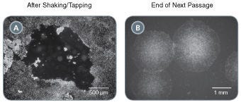
Figure 4. Select Putative iPS Cell Clones
Easily isolate newly generated human iPS cell colonies with ReLeSR™ by selectively detaching undifferentiated cells and leaving non reprogrammed cells behind. (A) A TeSR™-E7™ reprogramming culture which has been treated with ReLeSR™ to detach the putative iPS cell colony, leaving the non-reprogrammed and differentiated cells behind. (B) Cultures contain a high proportion of undifferentiated cells by the end of the first passage.
This product is designed for use in the following research area(s) as part of the highlighted workflow stage(s). Explore these workflows to learn more about the other products we offer to support each research area.
Pluripotent Stem Cell-Derived Cerebral Organoids Reveal Human Oligodendrogenesis with Dorsal and Ventral Origins. H. Kim et al. Stem cell reports 2019
Abstract
The process of oligodendrogenesis has been relatively well delineated in the rodent brain. However, it remains unknown whether analogous developmental processes are manifested in the human brain. Here we report oligodendrogenesis in forebrain organoids, generated by using OLIG2-GFP knockin human pluripotent stem cell (hPSC) reporter lines. OLIG2/GFP exhibits distinct temporal expression patterns in ventral forebrain organoids (VFOs) versus dorsal forebrain organoids (DFOs). Interestingly, oligodendrogenesis can be induced in both VFOs and DFOs after neuronal maturation. Assembling VFOs and DFOs to generate fused forebrain organoids (FFOs) promotes oligodendroglia maturation. Furthermore, dorsally derived oligodendroglial cells outcompete ventrally derived oligodendroglia and become dominant in FFOs after long-term culture. Thus, our organoid models reveal human oligodendrogenesis with ventral and dorsal origins. These models will serve to study the phenotypic and functional differences between human ventrally and dorsally derived oligodendroglia and to reveal mechanisms of diseases associated with cortical myelin defects.
Disruption of GRIN2B Impairs Differentiation in Human Neurons. S. Bell et al. Stem cell reports 2018 JUL
Abstract
Heterozygous loss-of-function mutations in GRIN2B, a subunit of the NMDA receptor, cause intellectual disability and language impairment. We developed clonal models of GRIN2B deletion and loss-of-function mutations in a region coding for the glutamate binding domain in human cells and generated neurons from a patient harboring a missense mutation in the same domain. Transcriptome analysis revealed extensive increases in genes associated with cell proliferation and decreases in genes associated with neuron differentiation, a result supported by extensive protein analyses. Using electrophysiology and calcium imaging, we demonstrate that NMDA receptors are present on neural progenitor cells and that human mutations in GRIN2B can impair calcium influx and membrane depolarization even in a presumed undifferentiated cell state, highlighting an important role for non-synaptic NMDA receptors. It may be this function, in part, which underlies the neurological disease observed in patients with GRIN2B mutations.
Intrinsic Immunity Shapes Viral Resistance of Stem Cells. Wu X et al. Cell 2018 JAN
Abstract
Stem cells are highly resistant to viral infection compared to their differentiated progeny; however, the mechanism is mysterious. Here, we analyzed gene expression in mammalian stem cells and cells at various stages of differentiation. We find that, conserved across species, stem cells express a subset of genes previously classified as interferon (IFN) stimulated genes (ISGs) but that expression is intrinsic, as stem cells are refractory to interferon. This intrinsic ISG expression varies in a cell-type-specific manner, and many ISGs decrease upon differentiation, at which time cells become IFN responsive, allowing induction of a broad spectrum of ISGs by IFN signaling. Importantly, we show that intrinsically expressed ISGs protect stem cells against viral infection. We demonstrate the in vivo importance of intrinsic ISG expression for protecting stem cells and their differentiation potential during viral infection. These findings have intriguing implications for understanding stem cell biology and the evolution of pathogen resistance.
Thank you for your interest in IntestiCult™ Organoid Growth Medium (Human). Please provide us with your contact information and your local representative will contact you with a customized quote. Where appropriate, they
can also assist you with a(n):
Estimated delivery time for your area
Product sample or exclusive offer
In-lab demonstration
By submitting this form, you are providing your consent to STEMCELL Technologies Canada Inc. and its subsidiaries and affiliates (“STEMCELL”) to collect and use your information, and send you newsletters and emails in accordance with our
privacy policy. Please contact us with any questions that you may have. You can unsubscribe or change your email preferences at any time.
THIS PRODUCT IS MANUFACTURED AND TESTED FOLLOWING RELEVANT CGMPs UNDER A CERTIFIED QUALITY MANAGEMENT SYSTEM. PRODUCT IS FOR FURTHER MANUFACTURING OR RESEARCH USE. NOT INTENDED FOR HUMAN OR ANIMAL DIAGNOSTIC OR THERAPEUTIC USES UNLESS OTHERWISE STATED. FOR ADDITIONAL INFORMATION ON QUALITY AT STEMCELL, REFER TO WWW.STEMCELL.COM/COMPLIANCE