产品号 #05702_C
小鼠和大鼠神经干细胞及祖细胞扩增培养基套装
Cell culture supplement
Basic fibroblast growth factor

Epidermal growth factor
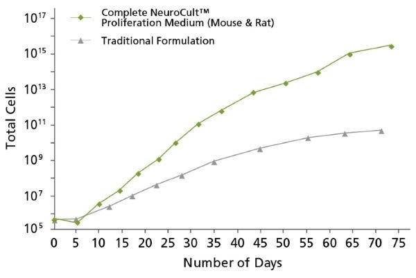
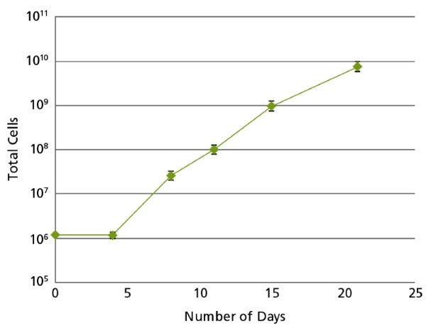
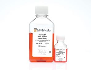
NeuroCult™增殖套装(小鼠和大鼠)是一种标准化的无血清培养基及添加剂套装,用于培养来自正常组织或肿瘤样本的小鼠和大鼠神经干细胞及祖细胞。在添加适当细胞因子后,NeuroCult™扩增套装(小鼠和大鼠)经过优化,可长时间维持培养小鼠和大鼠神经干细胞,且不会丧失其自我更新、扩增或分化潜能。
注意:配制NeuroCult™完全扩增培养基时,需添加人重组EGF(产品编号#78006.1)。培养来自成年小鼠或大鼠的细胞时,还需添加人重组bFGF(产品编号#78003.1)和肝素溶液(产品编号#07980)。
Subtype
Specialized Media
Cell Type
Brain Tumor Stem Cells, Neural Stem and Progenitor Cells
Species
Mouse, Rat
Application
Cell Culture, Colony Assay, Expansion, Functional Assay, Spheroid Culture, Toxicity Assay
Brand
NeuroCult
Area of Interest
Cancer, Disease Modeling, Neuroscience, Stem Cell Biology
Formulation Category
Serum-Free
Find supporting information and directions for use in the Product Information Sheet or explore additional protocols below.
This product is designed for use in the following research area(s) as part of the highlighted workflow stage(s). Explore these workflows to learn more about the other products we offer to support each research area.
| Species | Mouse, Rat |
|---|---|
| Formulation Category | Serum-Free |
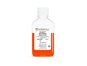
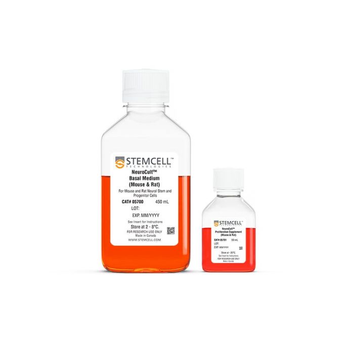
培养小鼠和大鼠神经干和祖细胞的基础培养基
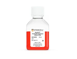
补充小鼠和大鼠神经干细胞和祖细胞的扩增
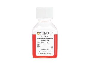
小鼠和大鼠神经干细胞和祖细胞分化的补充物
小鼠和大鼠神经干细胞和祖细胞分化培养基试剂盒