产品号 #06030_C
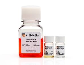
建立和维持小鼠肝祖类器官的细胞培养基
Rinsing solution for cultureware to prevent cell adhesion
Cell dissociation reagent
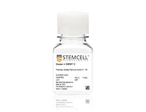
1 U/mL dispase in DMEM/F-12
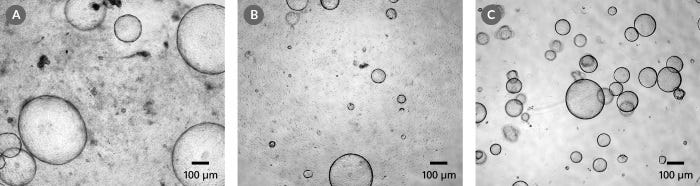
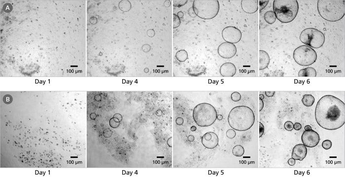
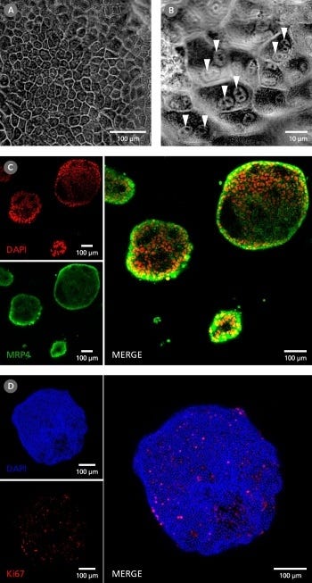
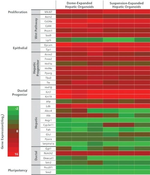
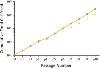
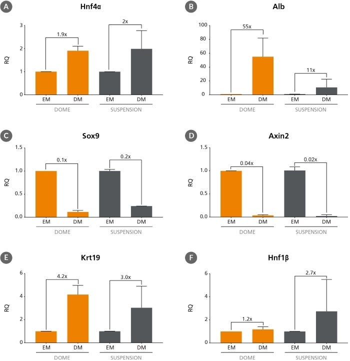
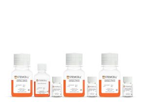
hepatcult™类器官生长培养基(小鼠)是一种无血清的细胞培养基,用于建立和维持小鼠肝祖类器官。这些类器官,或“迷你肝脏”,为研究肝干和祖细胞提供了体外器官型培养系统。在hepatcult™中生长的类器官具有表达标记肝干和祖细胞(PROM1, AXIN2, SOX9和CD44),导管(KRT19和HNF1b)和肝细胞(HNF4a, AFP)基因的上皮。肝类器官可以每4 - 7天传代一次,可以冷冻保存,并为下游分化做好准备。
hepatcult™支持嵌入康宁®Matrigel®圆顶或稀释Matrigel®悬浊液中的小鼠肝类器官培养。类器官培养可以方便地在生理相关系统中对肝上皮进行体外表征,并减少对动物使用的需求。
如果您打算将此产品用于商业用途,请通过www.huborganoids.nl获取商业用途许可证或HUB许可的相关说明。
Subtype
Specialized Media
Cell Type
Hepatic Cells
Species
Mouse
Application
Cell Culture, Expansion, Maintenance, Organoid Culture
Brand
HepatiCult
Area of Interest
Cancer, Disease Modeling, Drug Discovery and Toxicity Testing, Epithelial Cell Biology, Stem Cell Biology
Formulation Category
Serum-Free
Find supporting information and directions for use in the Product Information Sheet or explore additional protocols below.
This product is designed for use in the following research area(s) as part of the highlighted workflow stage(s). Explore these workflows to learn more about the other products we offer to support each research area.
| Species | Mouse |
|---|---|
| Formulation Category | Serum-Free |
建立和维持小鼠肠道类器官的细胞培养基
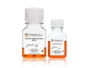
建立和维持人肠道类器官的细胞培养基
用于人肝脏类器官生成、生长和分化的培养基试剂盒