HepatiCult™ 类器官生长培养基 (小鼠)

用于建立和维持小鼠肝祖类器官的培养基

产品号 #(选择产品)

产品号 #06030_C

用于建立和维持小鼠肝祖类器官的培养基

产品优势

  • 便捷的体外系统,可在4-5天内生成类器官;
  • 步骤清晰的实验方案,无需损伤模型、手动挑选导管或细胞分选;
  • 简单的两组分形式;无血清且成分明确;
  • 灵活的培养方案,可以胶滴的形式或悬浮培养的形式生成并维持类器官的生长

产品组分包括

  • HepatiCult™ 类器官生长基础培养基(小鼠),95 mL
  • HepatiCult™ 类器官生长补充剂(小鼠), 5 mL
Interested in trying STEMCELL's organoid products for your liver research? Fill out this form to request information about introductory offers.

总览

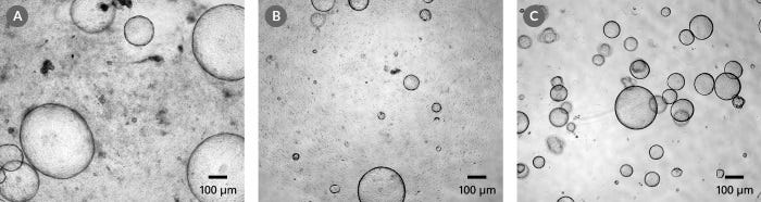
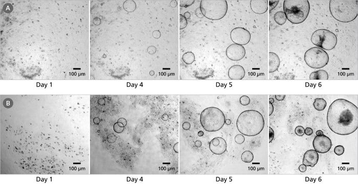
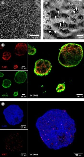
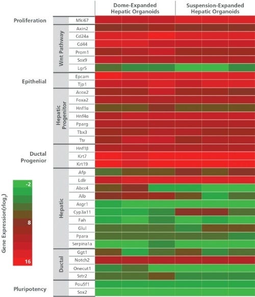
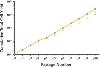
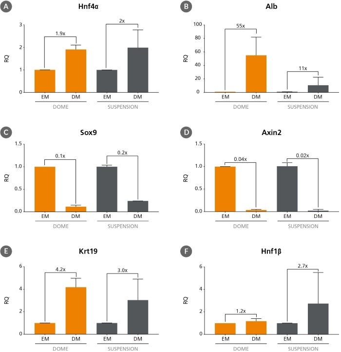
HepatiCult™ 类器官生长培养基 (小鼠)是一款无血清的成分明确的培养基,用于小鼠肝脏祖细胞类器官的建立和维持。这些类器官或称“迷你肝脏”,提供了一个体外的器官型培养系统,用于研究肝脏干细胞和祖细胞。在HepatiCult™培养基中生长的类器官具有表达标记肝脏干细胞和祖细胞(PROM1、AXIN2、SOX9和CD44)、导管细胞(KRT19和HNF1b)以及肝细胞(HNF4a、AFP)基因的上皮。肝类器官可以每4 - 7天传代一次,可进行冷冻保存,也可进行下游分化。

HepatiCult™支持将小鼠肝类器官在Corning® Matrigel®的胶滴中,或在稀释的Matrigel®悬液中进行培养。类器官培养提供了一种便捷的体外方法,可在生理相关的体系中对肝脏上皮进行表征,同时减少对动物的使用。

如果您打算将本产品用于商业目的,请通过www.huborganoids.nl与HUB联系,以获取商业用途许可或HUB许可相关的说明。

亚型
专用培养基
 
细胞类型
肝细胞
 
种属
小鼠
 
应用
细胞培养,扩增,培养,类器官培养
 
品牌
HepatiCult
 
研究领域
癌症,疾病建模,药物发现和毒理检测,上皮细胞研究,干细胞生物学
 
制剂类别
无血清
 

Data Figures

Figure 1. Mouse Hepatic Progenitor Organoids can be Initiated from a Variety of Starting Materials

HepatiCult™ Organoid Growth Medium (Mouse) enables the initiation of hepatic progenitor organoids from (A) duct fragments, (B) single cells or (C) cryopreserved organoids. All organoids were grown in Matrigel® domes and imaged on day 7 of primary culture or the first passage post thaw (cryopreserved organoids).

Figure 2. Hepatic Organoids can be Grown in Matrigel® Domes or as a Dilute Matrigel® Suspension

Hepatic progenitor organoids were cultured from freshly isolated mouse hepatic duct fragments in HepatiCult™ Organoid Growth Medium (Mouse) and plated (A) in Matrigel® domes or (B) as a dilute Matrigel® suspension. Organoids grown in either culture condition are ready for passage within 4-7 days.

Figure 3. Organoids grown in HepatiCult™ Organoid Growth Medium (Mouse) Display Some Characteristics Typical of the Mature Hepatic Epithelium

(A) Hepatic progenitor organoids exhibit the polygonal morphology typical of the hepatic epithelium. (B) Hepatic progenitors cultured as organoids show binucleation (arrows), a feature common of mature hepatocytes. (C) Immunocytochemistry analysis shows localization of MRP4 (green), a membrane-bound, unidirectional efflux transporter, along the exterior of the organoids and DAPI (red) localized to the cellular nuclei. This indicates cellular polarization of the organoids with the basolateral surface of the epithelium distal from the lumen. (D) Hepatic organoids contain an actively dividing progenitor population, seen through the expression of Ki67 (red). Cell nuclei are stained with DAPI (blue).

Figure 4. Analysis of Organoid Gene Expression when Grown in Matrigel® Domes and as a Dilute Matrigel® Suspension

Analysis of marker expression by RNA-seq shows organoids grown in HepatiCult™ Organoid Growth Medium (Mouse) in either Matrigel® domes or in a dilute Matrigel® suspension express markers associated with hepatic stem and progenitor cells. The organoids also express low levels of genes associated with mature hepatic cell types, including cholangiocytes and hepatocytes. Columns represent biological replicates at passages ranging from passage 1 - 40.

Figure 5. Expansion of Organoids grown in HepatiCult™ Organoid Growth Medium (Mouse)

Organoids cultured with HepatiCult™ Organoid Growth Medium (Mouse) show efficient growth over passaging. Cultures were split with an average split ratio of 1:25 at each passage.

Figure 6. Differentiation of Hepatic Progenitor Organoids

Hepatic progenitor organoids grown in HepatiCult™ Organoid Growth Medium (Mouse) (EM) can be differentiated to resemble more mature cell types when switched to a differentiation medium (DM)1,2. After switching to published differentiation medium hepatic organoids show the upregulation of mature hepatic markers such as (A) Hnf4α and (B) Alb, downregulation of hepatic stem cell and progenitor markers (C) Sox9 and (D) Axin2 and upregulation of ductal markers (E) Krt19 and (F) Hnf1β. Relative quantification (RQ) for each marker is reported relative to 18S and TBP housekeeping genes and normalized relative to hepatic progenitor organoids grown in HepatiCult™ Organoid Growth Medium (Mouse) and cultured in Matrigel® domes.

Protocols and Documentation

Find supporting information and directions for use in the Product Information Sheet or explore additional protocols below.

Document Type
Product Name
Catalog #
Lot #
Language
Catalog #
06030
Lot #
All
Language
English
Document Type
Safety Data Sheet 1
Catalog #
06030
Lot #
All
Language
English
Document Type
Safety Data Sheet 2
Catalog #
06030
Lot #
All
Language
English

Applications

This product is designed for use in the following research area(s) as part of the highlighted workflow stage(s). Explore these workflows to learn more about the other products we offer to support each research area.

Research Area
Workflow Stages
Workflow Stages for Organoids

Resources and Publications

Educational Materials (20)

Publications (2)

The CellRaft AIR? system: A novel system enabling organoid imaging, identification, and isolation A. Stern et al. SLAS Discovery 2022 4

Abstract

Three-dimensional (3D) culture systems have been developed that can re-capitulate organ level responses, simulate compound diffusion through complex structures, and assess cellular heterogeneity of tissues, making them attractive models for advanced in vitro research and discovery. Organoids are a unique subtype of 3D cell culture that are grown from stem cells, are self-organizing, and closely replicate in vivo pathophysiology. Organoids have been used to understand tissue development, model diseases, test drug sensitivity and toxicity, and advance regenerative medicine. However, traditional organoid culture methods are inadequate because they are low throughput and ill-suited for single organoid imaging, phenotypic assessment, and isolation from heterogenous organoid populations. To address these bottlenecks, we have adapted our tissue culture consumable and instrumentation to enable automated imaging, identification, and isolation of individual organoids. Organoids grown on the 3D CytoSort? Array can be reliably tracked, imaged, and phenotypically analyzed using brightfield and fluorescent microscopy as they grow over time, then released and transferred fully intact for use in downstream applications. Using mouse hepatic and pancreatic organoids, we have demonstrated the use of this technology for single-organoid imaging, clonal organoid generation, parent organoid subcloning, and single-organoid RNA extraction for downstream gene expression or transcriptomic analysis. The results validate the ability of the CellRaft AIR? System to facilitate efficient, user-friendly, and automated workflows broadly applicable to organoid research by overcoming several pain points: 1) single organoid time-course imaging and phenotypic assessment, 2) establishment of single cell-derived organoids, and 3) isolation and retrieval of single organoids for downstream applications.
Modulation of the extrinsic cell death signaling pathway by viral Flip induces acute-death mediated liver failure. M. Bittel et al. Cell death {\&} disease 2019

Abstract

During viral infections viruses express molecules that interfere with the host-cell death machinery and thus inhibit cell death responses. For example the viral FLIP (vFLIP) encoded by Kaposi's sarcoma-associated herpesvirus interacts and inhibits the central cell death effector, Caspase-8. In order to analyze the impact of anti-apoptotic viral proteins, like vFlip, on liver physiology in vivo, mice expressing vFlip constitutively in hepatocytes (vFlipAlbCre+) were generated. Transgenic expression of vFlip caused severe liver tissue injury accompanied by massive hepatocellular necrosis and inflammation that finally culminated in early postnatal death of mice. On a molecular level, hepatocellular death was mediated by RIPK1-MLKL necroptosis driven by an autocrine TNF production. The loss of hepatocytes was accompanied by impaired bile acid production and disruption of the bile duct structure with impact on the liver-gut axis. Notably, embryonic development and tissue homeostasis were unaffected by vFlip expression. In summary our data uncovered that transgenic expression of vFlip can cause severe liver injury in mice, culminating in multiple organ insufficiency and death. These results demonstrate that viral cell death regulatory molecules exhibit different facets of activities beyond the inhibition of cell death that may merit more sophisticated in vitro and in vivo analysis.

更多信息

更多信息
Species Mouse
Formulation Category Serum-Free
Legal Statement: This product was developed under a license to intellectual property owned by Hubrecht Organoid Technology (HUB). This product is sold for research use only. Purchase of this product does not include the right to use it for drug screening aiming for commercial gain, equipment validation, biobanking, or for other commercial purposes. Purchasers wishing to use the product for purposes other than basic research use should contact HUB at www.huborganoids.nl to obtain a further license. Purchasers may apply for a License from HUB, which will not be unreasonably withheld by HUB. PRODUCTS ARE FOR RESEARCH USE ONLY AND NOT INTENDED FOR HUMAN OR ANIMAL DIAGNOSTIC OR THERAPEUTIC USES UNLESS OTHERWISE STATED. FOR ADDITIONAL INFORMATION ON QUALITY AT STEMCELL, REFER TO WWW.STEMCELL.COM/COMPLIANCE.
版权 © 2025 STEMCELL Technologies 技术有限公司。保留所有权利。