MesenCult™-ACF Plus培养试剂盒

用于人间充质干细胞的无动物成分培养基

产品号 #(选择产品)

产品号 #05448_C

用于人间充质干细胞的无动物成分培养基

产品优势

  • 无动物成分配方提升实验可重复性。
  • 相较于含血清培养基,具备更优异的细胞扩增效果。
  • 培养的MSC在早期和晚期传代中均保持强劲的扩增能力及三系分化潜能。
  • 支持直接从人类原代组织中衍生MSC。

产品组分包括

  • MesenCult™ -ACF Plus基础培养基(500 mL)
  • MesenCult™-ACF Plus 500X 补充剂(1 mL)
  • 无动物成分细胞贴附基质 (1 mL)

概述

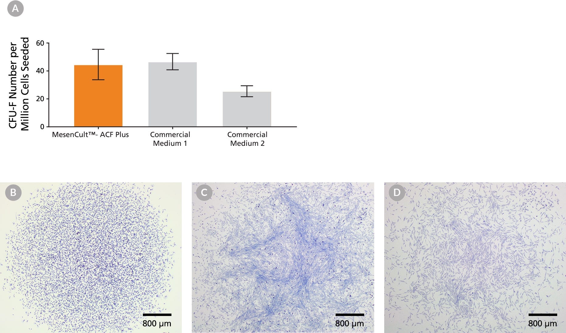
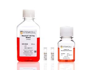
使用这款无动物成分(ACF)且不含细胞外囊泡(EV)的培养基,可降低人间充质基质细胞(MSC,亦称间充质干细胞)培养的差异性,提升实验可重复性。MesenCult™-ACF Plus培养试剂盒经过优化,无需血清即可从骨髓或脂肪组织等多种来源衍生人类MSC。

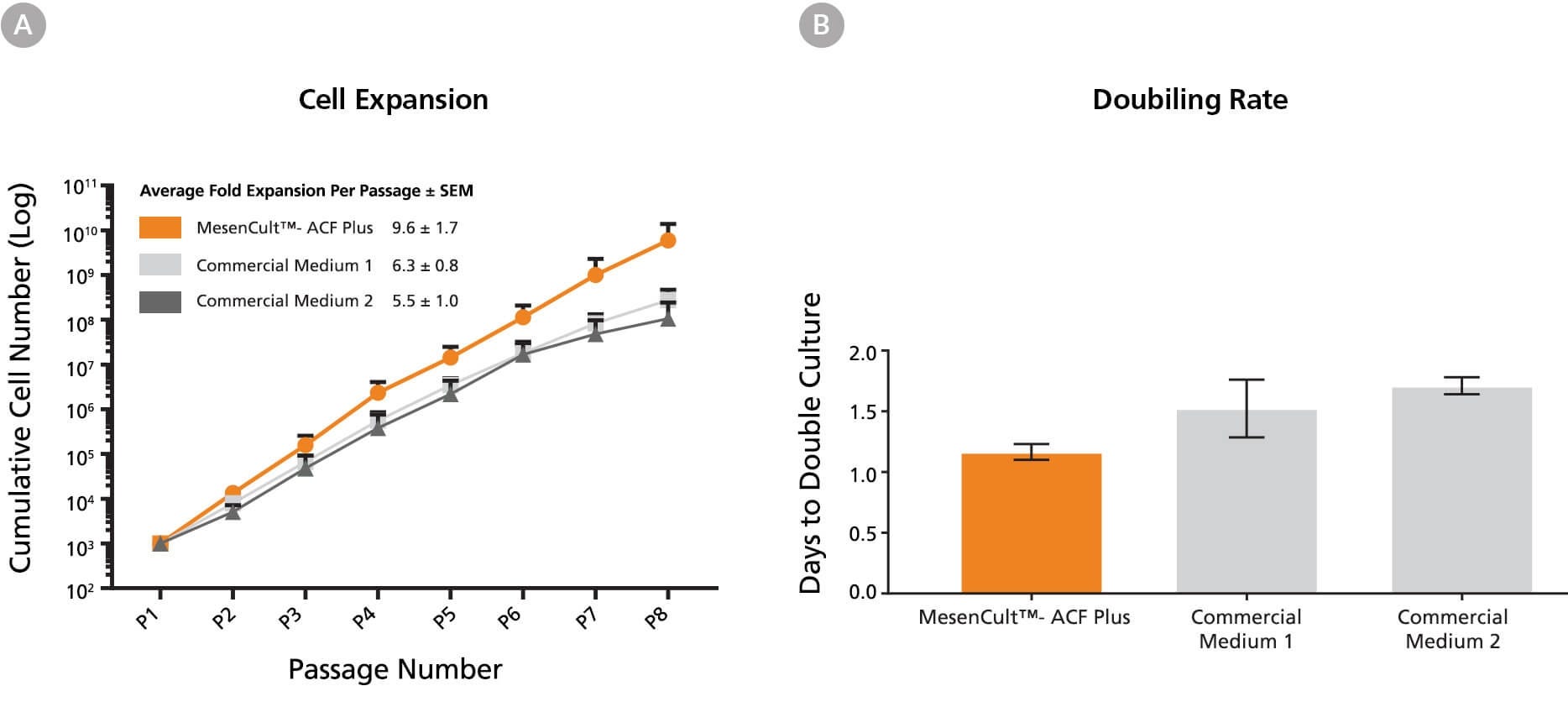
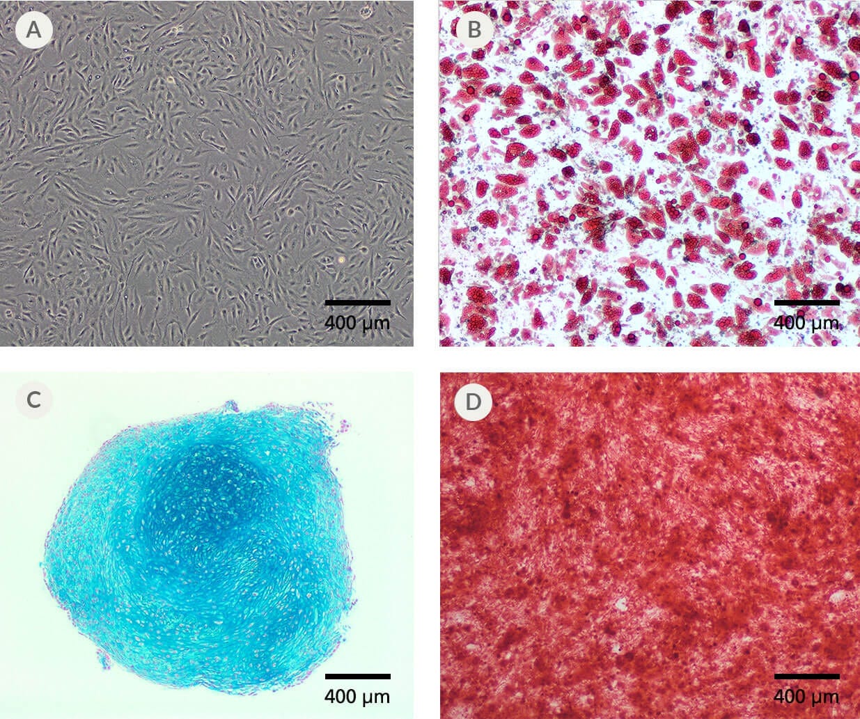
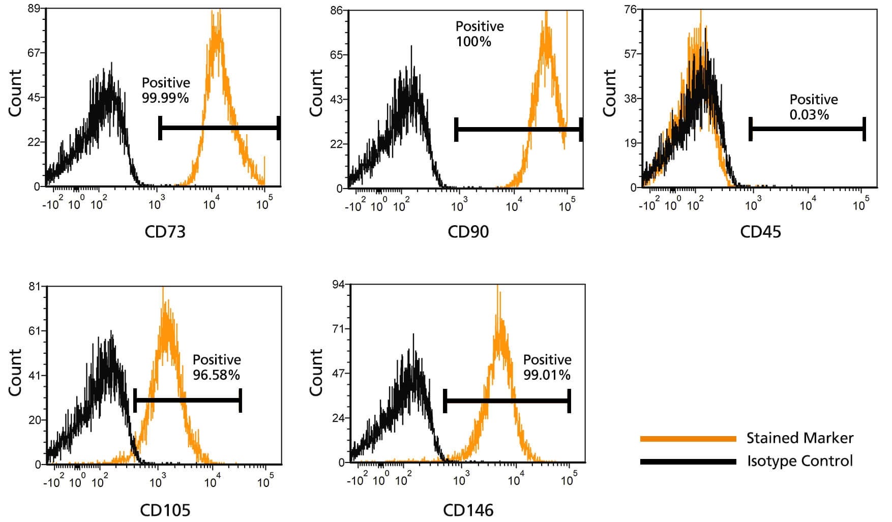
与含血清或EV去除的含血清培养基相比,使用本试剂盒培养的MSC扩增效率更高(增殖速率与累计细胞总量),且功能不受影响。培养细胞表达典型的MSC表面标志物,并保持强劲的扩增速率和三系分化能力。

本试剂盒是完整ACF产品线的一部分——涵盖MSC的衍生、扩增、冻存;以及将人多能干细胞分化为间充质祖细胞——专为高效稳定的MSC培养而优化。

为实现无动物成分的优化的细胞冻存目的,推荐使用MesenCult™-ACF冻存培养基冻存先前在MesenCult™培养基(包括MesenCult™-ACF Plus)中培养的人类MSC。有关相关产品的完整列表,包括可用的分化培养基,请访问 our MSC area of interest page,或通过 techsupport@stemcell.com 联系我们获取。

注意:MesenCult™-ACF Plus完全培养基必须添加L-谷氨酰胺。配制完全培养基所需的培养基与补充剂(不含基质)亦可单独购买,即MesenCult™-ACF Plus培养基。

CollPlant是细胞贴附基质中重组人胶原蛋白(rhCollagen)组分的生产商。

本产品仅限研究使用。如有任何临床或商业应用需求,请联系STEMCELL公司。

Subtype
Specialized Media
 
Cell Type
Mesenchymal Cells, PSC-Derived, Mesenchymal Stem and Progenitor Cells
 
Species
Human
 
Application
Cell Culture, Expansion, Maintenance
 
Brand
MesenCult
 
Area of Interest
Extracellular Vesicle Research, Stem Cell Biology
 
Formulation Category
Animal Component-Free, Serum-Free
 

Data Figures

Figure 1. CFU-F Assay of Human BM-Derived MSCs Expanded in MesenCult™-ACF Plus Medium and Commercial Media.
(A) An average of 45 CFU-Fs per million cells were observed when BM mononuclear cells were seeded in MesenCult™-ACF Plus (n = 4). An average of 47 and 25 CFU-Fs per million cells were observed when cells were seeded in Commercial Medium 1 (n = 3) and Medium 2 (n = 4), respectively. Vertical lines indicate Standard Error of Mean (SEM). Representative image of CFU-F colonies expanded in (B) MesenCult™-ACF Plus Medium (9 days of culture), (C) Commercial Medium 1 (10 days of culture) and (D) Commercial Medium 2 (10 days of culture). Commercial Medium 1 and Medium 2 were supplemented with 2.5% human AB serum to derive MSCs from BM, as per their protocols for derivation. No addition of serum is required when using MesenCult™-ACF Plus Medium.

Figure 2. Human BM-Derived MSCs Cultured in MesenCult™-ACF Plus Medium Expand Faster than MSCs Cultured in Commercial Xeno-Free and Serum-Free Media.
(A) A greater number of BM-derived MSCs were generated per passage using MesenCult™-ACF Plus Medium (n=4) compared to Commercial Medium 1 (n=3) and Commercial Medium 2 (n=2). (B) Rates of BM-derived MSC expansion were compared between MesenCult™-ACF Plus Medium, Commercial Medium 1, and Commercial Medium 2. The time required to double the number of MSCs using MesenCult™ -ACF Plus Medium (n=4) was shorter than when MSCs were cultured in Commercial Medium 1 (n=3) and Commercial Medium 2 (n=4). Vertical lines indicate Standard Error of Mean (SEM).

Figure 3. Human BM-Derived MSCs Expanded in MesenCult™-ACF Plus Medium Display Multi-Lineage Differentiation Potential.
(A) Human BM-derived MSCs expanded in MesenCult™-ACF Plus Medium differentiated into (B) adipocytes (Oil Red O staining; passage 5), (C) chondrocytes (Alcian Blue staining; passage 4) and (D) osteoblasts (Alizarin Red S staining; passage 5).

Figure 4. Flow Cytometric Analysis of MSCs Cultured in MesenCult™-ACF Plus Medium.
BM-derived MSCs were cultured and expanded in MesenCult™-ACF Plus Medium. At passage 8 MSCs were stained for mesenchymal surface markers (CD73, CD90, CD105,), pericyte marker (CD146) and hematopoietic marker (CD45). MSCs expressed high levels of CD73, CD90, CD105 and CD146 and lacked expression of CD45.

Protocols and Documentation

Find supporting information and directions for use in the Product Information Sheet or explore additional protocols below.

Document Type
Product Name
Catalog #
Lot #
Language
Catalog #
05448
Lot #
All
Language
English
Document Type
Safety Data Sheet 1
Catalog #
05448
Lot #
All
Language
English
Document Type
Safety Data Sheet 2
Catalog #
05448
Lot #
All
Language
English
Document Type
Safety Data Sheet 3
Catalog #
05448
Lot #
All
Language
English

Applications

This product is designed for use in the following research area(s) as part of the highlighted workflow stage(s). Explore these workflows to learn more about the other products we offer to support each research area.

Resources and Publications

Educational Materials (13)

Publications (8)

CD81 Controls Beige Fat Progenitor Cell Growth and Energy Balance via FAK Signaling. Y. Oguri et al. Cell 2020 jun

Abstract

Adipose tissues dynamically remodel their cellular composition in response to external cues by stimulating beige adipocyte biogenesis; however, the developmental origin and pathways regulating this process remain insufficiently understood owing to adipose tissue heterogeneity. Here, we employed single-cell RNA-seq and identified a unique subset of adipocyte progenitor cells (APCs) that possessed the cell-intrinsic plasticity to give rise to beige fat. This beige APC population is proliferative and marked by cell-surface proteins, including PDGFR$\alpha$, Sca1, and CD81. Notably, CD81 is not only a beige APC marker but also required for de novo beige fat biogenesis following cold exposure. CD81 forms a complex with $\alpha$V/$\beta$1 and $\alpha$V/$\beta$5 integrins and mediates the activation of integrin-FAK signaling in response to irisin. Importantly, CD81 loss causes diet-induced obesity, insulin resistance, and adipose tissue inflammation. These results suggest that CD81 functions as a key sensor of external inputs and controls beige APC proliferation and whole-body energy homeostasis.
Despite mutation acquisition in hematopoietic stem cells, JMML-propagating cells are not always restricted to this compartment. A. Caye et al. Leukemia 2020 jun

Abstract

Juvenile myelomonocytic leukemia (JMML) is a rare aggressive myelodysplastic/myeloproliferative neoplasm of early childhood, initiated by RAS-activating mutations. Genomic analyses have recently described JMML mutational landscape; however, the nature of JMML-propagating cells (JMML-PCs) and the clonal architecture of the disease remained until now elusive. Combining genomic (exome, RNA-seq), Colony forming assay and xenograft studies, we detect the presence of JMML-PCs that faithfully reproduce JMML features including the complex/nonlinear organization of dominant/minor clones, both at diagnosis and relapse. Further integrated analysis also reveals that although the mutations are acquired in hematopoietic stem cells, JMML-PCs are not always restricted to this compartment, highlighting the heterogeneity of the disease during the initiation steps. We show that the hematopoietic stem/progenitor cell phenotype is globally maintained in JMML despite overexpression of CD90/THY-1 in a subset of patients. This study shed new lights into the ontogeny of JMML, and the identity of JMML-PCs, and provides robust models to monitor the disease and test novel therapeutic approaches.
Precision installation of a highly efficient suicide gene safety switch in human induced pluripotent stem cells. Z.-D. Shi et al. Stem cells translational medicine 2020 jul

Abstract

Human pluripotent stem cells including induced pluripotent stem cells (iPSCs) and embryonic stem cells hold great promise for cell-based therapies, but safety concerns that complicate consideration for routine clinical use remain. Installing a safety switch" based on the inducible caspase-9 (iCASP9) suicide gene system should offer added control over undesirable cell replication or activity. Previous studies utilized lentiviral vectors to integrate the iCASP9 system into T cells and iPSCs. This method results in random genomic insertion of the suicide switch and inefficient killing of the cells after the switch is "turned on" with a small molecule (eg AP1903). To improve the safety and efficiency of the iCASP9 system for use in iPSC-based therapy we precisely installed the system into a genomic safe harbor the AAVS1 locus in the PPP1R12C gene. We then evaluated the efficiencies of different promoters to drive iCASP9 expression in human iPSCs. We report that the commonly used EF1$\alpha$ promoter is silenced in iPSCs and that the endogenous promoter of the PPP1R12C gene is not strong enough to drive high levels of iCASP9 expression. However the CAG promoter induces strong and stable iCASP9 expression in iPSCs and activation of this system with AP1903 leads to rapid killing and complete elimination of iPSCs and their derivatives including MSCs and chondrocytes in vitro. Furthermore iPSC-derived teratomas shrank dramatically or were completely eliminated after administration of AP1903 in mice. Our data suggest significant improvements on existing iCASP9 suicide switch technologies and may serve as a guide to other groups seeking to improve the safety of stem cell-based therapies."

更多信息

更多信息
Species Human
Formulation Category Animal Component-Free, Serum-Free
PRODUCTS ARE FOR RESEARCH USE ONLY AND NOT INTENDED FOR HUMAN OR ANIMAL DIAGNOSTIC OR THERAPEUTIC USES UNLESS OTHERWISE STATED. FOR ADDITIONAL INFORMATION ON QUALITY AT STEMCELL, REFER TO WWW.STEMCELL.COM/COMPLIANCE.
版权 © 2025 STEMCELL Technologies 技术有限公司。保留所有权利。