MesenCult™ 脂肪分化试剂盒 (人)

人MSCs向脂肪细胞分化的培养基

产品号 #(选择产品)

产品号 #05412_C

人MSCs向脂肪细胞分化的培养基

产品优势

  • 高效和通用的人MSC向脂肪细胞分化培养基;
  • 为先前在含血清或无血清培养基(如MesenCult™-ACF Plus 培养基)中培养的骨髓和脂肪组织来源的人MSCs的分化而优化;
  • 先前在含血小板裂解物培养基中培养的人MSCs兼容

产品组分包括

  • MesenCult™ MSC 脂肪分化基础培养基 (人), 225 mL
  • MesenCult™ 10X 脂肪分化补充剂 (人), 25 mL
  • MesenCult™ 500X 脂肪分化补充剂 (人), 0.5 mL

总览

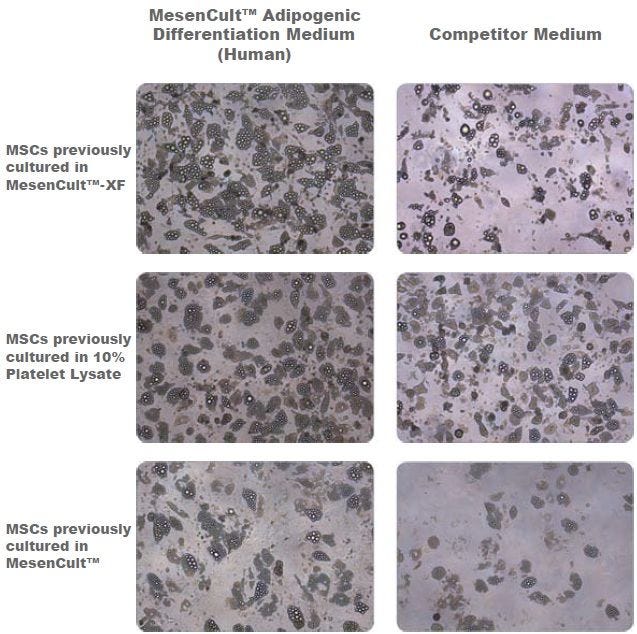
MesenCult™ 脂肪分化试剂盒 (人) 是专门为原代人间充质间质细胞和多能干细胞来源的间充质祖细胞(也称为间充质干细胞或MSCs)在体外分化成脂肪生成谱系细胞而研发。该试剂盒适用于分化来源于人骨髓(Bone marrow),脂肪组织(Adipose tissue),脐带组织或多能干细胞的 MSCs的成脂分化。这些MSCs分化前可在无血清和无动物成分培养基(例如MesenCult™ACF Plus培养基[产品号 #05445]),含血清培养基(例如MesenCult™ 扩增试剂盒 (人)[产品号 #05411])或含血小板裂解物培养基(例如MesenCult™-hPL培养基试剂盒[产品号 #05439])中培养扩增。

亚型
专用培养基
 
细胞类型
脂肪细胞,间充质干/祖细胞
 
种属

 
应用
细胞培养,分化
 
品牌
MesenCult
 
研究领域
干细胞生物学
 

Data Figures

Adipogenic Differentiation of Human Bone Marrow-Derived MSCs

Figure 1. Adipogenic Differentiation of Human Bone Marrow-Derived MSCs

Adipogenic differentiation of human bone marrow-derived MSCs using MesenCult™ Adipogenic Differentiation Medium (Human) or a competitor medium. Prior to differentiation, MSCs were cultured for 2 passages in either serum- and xeno-free media (MesenCult™-XF or a 10% platelet lysate-based formulation) or serum-containing medium (MesenCult™) before undergoing differentiation. Even though differentation results are donor dependent, MesenCult™ Adipogenic Differentiation Medium (Human) consistently performed as well as, or better than the competitor medium. This trend was consistent for MSCs previously cultured in MesenCult™-XF, 10% Platelet Lysate or MesenCult™ medium.

Protocols and Documentation

Find supporting information and directions for use in the Product Information Sheet or explore additional protocols below.

Document Type
Product Name
Catalog #
Lot #
Language
Catalog #
05412
Lot #
All
Language
English
Document Type
Safety Data Sheet 1
Catalog #
05412
Lot #
All
Language
English
Document Type
Safety Data Sheet 2
Catalog #
05412
Lot #
All
Language
English
Document Type
Safety Data Sheet 3
Catalog #
05412
Lot #
All
Language
English
Document Type
Safety Data Sheet 4
Catalog #
05412
Lot #
All
Language
English

Applications

This product is designed for use in the following research area(s) as part of the highlighted workflow stage(s). Explore these workflows to learn more about the other products we offer to support each research area.

Resources and Publications

Educational Materials (6)

Publications (13)

Differential expression of drug resistance genes in CD146 positive dental pulp derived stem cells and CD146 negative fibroblasts. M. S. Tavangar et al. Clinical and experimental dental research 2020 may

Abstract

INTRODUCTION The stem cell portion of the dental pulp derived cultures (DPSCs) showed a higher resistance to cytotoxic effect of restorative dental materials compared to pulpal fibroblasts (DPFs). Here, we aimed to compare the expression of some drug resistant genes between these cells. METHODS AND MATERIALS To separate DPSCs from DPFs, we used magnetic cell sorting technique based on CD146 expression. To assess the stem cell properties, the positive and negative portions underwent colony forming assays and were induced to be differentiated into the adipocytes, osteoblasts, hepatocytes, and neural cells. Cell surface antigen panels were checked using immune fluorescence and flow-cytometry techniques. The mRNA expression of 14 ABC transporters including ABCA2, ABCB1, ABCB11, ABCC1, ABCC2, ABCC3, ABCC4, ABCC5-2, ABCC5-4,ABCC5-13, ABCC6, ABCC10, ABCC11, and ABCG2 genes was assessed, using quantitative RT-PCR technique. RESULTS Only the CD146 positive portion could be differentiated into the desired fates, and they formed higher colonies (16.7 ± 3.32 vs. 1.7 ± 1.67, p {\textless} .001). The cell surface antigen panels were the same, except for CD146 and STRO-1 markers which were expressed only in the positive portion. Among the ABC transporter genes studied, the positive portion showed a higher expression (approximately two-fold) of ABCA2, ABCC5-13, and ABCC5-2 genes. CONCLUSION Dental pulp stem cells which can be separated from dental pulp fibroblasts based on CD146 expression, express higher levels of some drug resistance genes which probably accounts for their features of more resistance to cytotoxic effects of some dental materials. This needs to be more validated in future.
In situ repair abilities of human umbilical cord-derived mesenchymal stem cells and autocrosslinked hyaluronic acid gel complex in rhesus monkeys with intrauterine adhesion. L. Wang et al. Science advances 2020 may

Abstract

Increasing occurrence of moderate to severe intrauterine adhesion (IUA) is seriously affecting the quality of human life. The aim of the study was to establish IUA models in nonhuman primates and to explore the dual repair effects of human umbilical cord-derived mesenchymal stem cells (huMSCs) loaded on autocrosslinked hyaluronic acid gel (HA-GEL) on endometrial damage and adhesion. Here, we recorded the menstrual cycle data in detail with uterine cavities observed and endometrial tissues detected after intervention, and the thicker endometria, decreased amount of fibrotic formation, increased number of endometrium glands, etc., suggested that both HA-GEL and huMSC/HA-GEL complexes could partially repair IUA caused by mechanical injury, but huMSC/HA-GEL complex transplantation had notable dual repair effects: a reliable antiadhesion property and the promotion of endometrial regeneration.
Intravenous administration of iPS-MSCSPIONs mobilized into CKD parenchyma and effectively preserved residual renal function in CKD rat. J.-J. Sheu et al. Journal of cellular and molecular medicine 2020 mar

Abstract

This study traced intravenously administered induced pluripotent stem cell (iPSC)-derived mesenchymal stem cells (MSC) and assessed the impact of iPSC-MSC on preserving renal function in SD rat after 5/6 nephrectomy. The results of in vitro study showed that FeraTrack™Direct contrast particles (ie intracellular magnetic labelling) in the iPSC-MSC (ie iPS-MSCSPIONs ) were clearly identified by Prussian blue stain. Adult-male SD rats (n = 40) were categorized into group 1 (SC), group 2 [SC + iPS-MSCSPIONs (1.0 × 10⁶ cells)/intravenous administration post-day-14 CKD procedure], group 3 (CKD), group 4 [CKD + iPS-MSCSPIONs (0.5 × 10⁶ cells)] and group 5 [CKD + iPS-MSCSPIONs (1.0 × 10⁶ cells)]. By day-15 after CKD induction, abdominal MRI demonstrated that iPS-MSCSPIONs were only in the CKD parenchyma of groups 4 and 5. By day 60, the creatinine level/ratio of urine protein to urine creatinine/kidney injury score (by haematoxylin and eosin stain)/fibrotic area (Masson's trichrome stain)/IF microscopic finding of kidney injury molecule-1 expression was lowest in groups 1 and 2, highest in group 3, and significantly higher in group 4 than in group 5, whereas IF microscopic findings of podocyte components (ZO-1/synaptopodin) and protein levels of anti-apoptosis ((Bad/Bcl-xL/Bcl-2) exhibited an opposite pattern to creatinine level among the five groups (all P {\textless} .0001). The protein expressions of cell-proliferation signals (PI3K/p-Akt/m-TOR, p-ERK1/2, FOXO1/GSK3$\beta$/p90RSK), apoptotic/DNA-damage (Bax/caspases8-10/cytosolic-mitochondria) and inflammatory (TNF-$\alpha$/TNFR1/TRAF2/NF-$\kappa$B) biomarkers displayed an identical pattern to creatinine level among the five groups (all P {\textless} .0001). The iPS-MSCSPIONs that were identified only in CKD parenchyma effectively protected the kidney against CKD injury.

更多信息

更多信息
Species Human
PRODUCTS ARE FOR RESEARCH USE ONLY AND NOT INTENDED FOR HUMAN OR ANIMAL DIAGNOSTIC OR THERAPEUTIC USES UNLESS OTHERWISE STATED. FOR ADDITIONAL INFORMATION ON QUALITY AT STEMCELL, REFER TO WWW.STEMCELL.COM/COMPLIANCE.
版权 © 2025 STEMCELL Technologies 技术有限公司。保留所有权利。