ImmunoCult™ XF 人T细胞扩增培养基

用于T细胞扩增的无血清和无异种成分培养基

产品号 #(选择产品)

产品号 #10981_C

用于T细胞扩增的无血清和无异种成分培养基

产品优势

  • 无需在培养基中添加血清;
  • 支持稳定的T细胞扩增,培养10 - 12天后仍具有较高的活率;
  • 扩增的T细胞再刺激时能够产生包括IFN-γ和IL-4在内的细胞因子;
  • 与ImmunoCult™人T细胞激活剂(产品号#10970或#10971)搭配使用,用于T细胞的无磁珠活化

总览

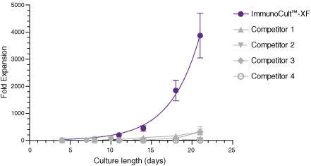
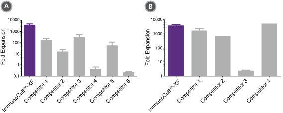
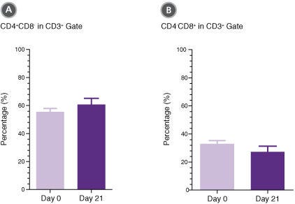
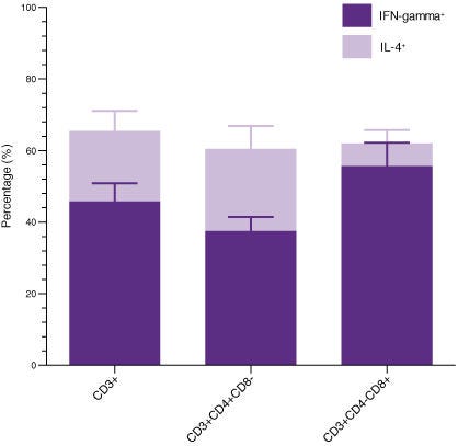
Immunocult™-XF T细胞扩增培养基是一种无血清和无异种成分培养基,用于体外培养和扩增外周血中分选的T细胞。适合T细胞的生长和扩增的重组细胞因子尚未添加到ImmunoCult™-XF T细胞扩增培养基中,以便用户灵活地制备满足其要求的完全培养基。
本产品仅用于为科研,如果您需要适合细胞治疗生产级别的试剂,推荐ImmunoCult™-XF(产品号#100-0956),该产品生产满足cGMP级相关规定,可用于临床应用。

亚型
专用培养基
 
细胞类型
T 细胞,T 细胞,CD4+,T 细胞,CD8+
 
种属
人,小鼠
 
应用
细胞培养,扩增
 
品牌
ImmunoCult
 
研究领域
免疫,细胞治疗开发
 
制剂类别
无血清,Xeno-Free
 

Data Figures

ImmunoCult™-XF T Cell Expansion Medium Supports Faster T Cell Expansion Than Other Serum-Free and Serum-Supplemented Media

Figure 1. ImmunoCult™-XF T Cell Expansion Medium Supports Faster T Cell Expansion Than Other Serum-Free and Serum-Supplemented Media

T cells were isolated from human peripheral blood samples using the EasySep™ Human T Cell Isolation Kit (Catalog #17951), stimulated with ImmunoCult™ Human CD3/CD28/CD2 T Cell Activator (Catalog #10970), and cultured in ImmunoCult™-XF T Cell Expansion Medium supplemented with rhIL-2. T cells were stimulated with ImmunoCult™ Human CD3/CD28/CD2 T Cell Activator on day 0 and every 7 to 8 days for the duration of the culture. T cells were analyzed on days 4, 7, 8, 10, 11, 14, 18, and 21 for fold expansion relative to the initial cell seeding density. Compared to all competitor media tested, ImmunoCult™-XF T Cell Expansion Medium showed significantly higher expansion of total T cells. Competitors 1 to 4 include, in no particular order, X-VIVO™ 15 (Lonza), AIM V® Medium (Life Tech), CellGro® DC Medium (CellGenix), and RPMI 1640 + serum. Each data point represents the mean fold expansion ± S.E.M. at the specified time points (p<0.05 for ImmunoCult™-XF versus all media for days 8, 11, 14, 18, and 21, tested using two-tailed, paired t-test with unequal variance, n = 6 to 19 donors). The average fold expansion of T cells in ImmunoCult™-XF T Cell Expansion Medium were 15-fold on day 7, 80-fold on day 10, 450-fold on day 14, and 4,000-fold on day 21.

ImmunoCult™-XF T Cell Expansion Medium Supports Greater T Cell Expansion Than Other Serum-Free and Serum-Supplemented Media

Figure 2. ImmunoCult™-XF T Cell Expansion Medium Supports Greater T Cell Expansion Than Other Serum-Free and Serum-Supplemented Media

T cells were isolated from human peripheral blood samples using the EasySep™ Human T Cell Isolation Kit (Catalog #17951), stimulated with ImmunoCult™ Human CD3/CD28/CD2 T Cell Activator (Catalog #10970), and cultured in (A) ImmunoCult™-XF T Cell Expansion Medium or serum-free competitor media with rhIL-2 in three replicate cultures per donor, or cultured in (B) ImmunoCult™-XF T Cell Expansion Medium or serum-supplemented competitor media with rhIL-2 in three replicate cultures per donor. T cells were stimulated with ImmunoCult™ Human CD3/CD28/CD2 T Cell Activator on day 0 and every 7 to 8 days for the duration of the culture. T cells were analyzed on day 21 for fold expansion relative to the initial cell seeding density.
(A) Compared to all serum-free competitor media tested, ImmunoCult™-XF T Cell Expansion Medium showed significantly higher expansion of total T cells. Competitors 1 to 6 represent serum-free competitor media, which include, in no particular order, X-VIVO™ 15 (Lonza), AIM V® Medium (Life Tech), CellGro® DC Medium (CellGenix), CTS™ OpTmizer™ T Cell Expansion SFM (Life Tech), TexMACS™ Medium (Miltenyi), and PRIME-XV® T Cell Expansion XSFM (Irvine Scientific). Each column with error bars represents the mean ± S.E.M. (p<5x10-13 for ImmunoCult™-XF T Cell Expansion Medium versus all other serum-free media, tested using the linear mixed effect model with linear regression, n = 4 to 19 donors).
(B) Compared to all serum-supplemented competitor media tested, ImmunoCult™-XF T Cell Expansion Medium showed similar or significantly higher expansion of total T cells. Competitors 1 to 4 represent serum-supplemented competitor media, which include, in no particular order, X-VIVO™ 15 + serum, CTS™ OpTmizer™ T Cell Expansion SFM + serum, RPMI 1640 + serum, and IMDM + serum. Each column with error bars represents the mean ± S.E.M. (p<0.0006 for ImmunoCult™-XF T Cell Expansion Medium versus all other serum-supplemented media except for Competitor 4, tested using the linear mixed effect model with linear regression, n = 1 to 19 donors).

T Cells Expanded in ImmunoCult™-XF T Cell Expansion Medium Show Similar Proportions of CD4+ and CD8+ Cells as T Cells at the Start of Culture

Figure 3. T Cells Expanded in ImmunoCult™-XF T Cell Expansion Medium Show Similar Proportions of CD4+ and CD8+ Cells as T Cells at the Start of Culture

T cells were isolated from human peripheral blood samples using the EasySep™ Human T Cell Isolation Kit (Catalog #17951), stimulated with ImmunoCult™ Human CD3/CD28/CD2 T Cell Activator (Catalog #10970), and cultured in ImmunoCult™-XF T Cell Expansion Medium supplemented with rhIL-2. T cells were stimulated with ImmunoCult™ Human CD3/CD28/CD2 T Cell Activator on day 0 and every 7 to 8 days for the duration of the culture. On day 0 and day 21, T cells were harvested and analyzed for (A) CD4+ and (B) CD8+ expression. Each column with error bars represents the mean ± S.E.M. (n = 24 donors for day 0 and n = 19 donors for day 21).

T Cells Expanded in ImmunoCult™-XF T Cell Expansion Medium Produce Intracellular IFN-gamma and IL-4

Figure 4. T Cells Expanded in ImmunoCult™-XF T Cell Expansion Medium Produce Intracellular IFN-gamma and IL-4

T cells were isolated from human peripheral blood samples using the EasySep™ Human T Cell Isolation Kit (Catalog #17951), stimulated with ImmunoCult™ Human CD3/CD28/CD2 T Cell Activator (Catalog #10970), and cultured in ImmunoCult™-XF T Cell Expansion Medium supplemented with rhIL-2. T cells were stimulated with ImmunoCult™ Human CD3/CD28/CD2 T Cell Activator on day 0 and every 7 to 8 days for the duration of the culture. On day 21, T cells were harvested and analyzed for intracellular IFN-gamma and IL-4 after stimulation with PMA and ionomycin for 4 hours and with Brefeldin A for 2 hours. The production of IFN-gamma and IL-4 in CD3+, CD3+CD4+CD8-, and CD3+CD4-CD8+ cells were determined. Each stacked column with error bars represents the mean ± S.E.M. (n = 9 donors).

Protocols and Documentation

Find supporting information and directions for use in the Product Information Sheet or explore additional protocols below.

Document Type
Product Name
Catalog #
Lot #
Language
Catalog #
10981
Lot #
All
Language
English
Document Type
Safety Data Sheet
Catalog #
10981
Lot #
All
Language
English

Applications

This product is designed for use in the following research area(s) as part of the highlighted workflow stage(s). Explore these workflows to learn more about the other products we offer to support each research area.

Resources and Publications

Educational Materials (31)

Publications (9)

Direct targeting of FOXP3 in Tregs with AZD8701, a novel antisense oligonucleotide to relieve immunosuppression in cancer. A. Revenko et al. Journal for immunotherapy of cancer 2022 apr

Abstract

BACKGROUND The Regulatory T cell (Treg) lineage is defined by the transcription factor FOXP3, which controls immune-suppressive gene expression profiles. Tregs are often recruited in high frequencies to the tumor microenvironment where they can suppress antitumor immunity. We hypothesized that pharmacological inhibition of FOXP3 by systemically delivered, unformulated constrained ethyl-modified antisense oligonucleotides could modulate the activity of Tregs and augment antitumor immunity providing therapeutic benefit in cancer models and potentially in man. METHODS We have identified murine Foxp3 antisense oligonucleotides (ASOs) and clinical candidate human FOXP3 ASO AZD8701. Pharmacology and biological effects of FOXP3 inhibitors on Treg function and antitumor immunity were tested in cultured Tregs and mouse syngeneic tumor models. Experiments were controlled by vehicle and non-targeting control ASO groups as well as by use of multiple independent FOXP3 ASOs. Statistical significance of biological effects was evaluated by one or two-way analysis of variance with multiple comparisons. RESULTS AZD8701 demonstrated a dose-dependent knockdown of FOXP3 in primary Tregs, reduction of suppressive function and efficient target downregulation in humanized mice at clinically relevant doses. Surrogate murine FOXP3 ASO, which efficiently downregulated Foxp3 messenger RNA and protein levels in primary Tregs, reduced Treg suppressive function in immune suppression assays in vitro. FOXP3 ASO promoted more than 70% reduction in FOXP3 levels in Tregs in vitro and in vivo, strongly modulated Treg effector molecules (eg, ICOS, CTLA-4, CD25 and 4-1BB), and augmented CD8+ T cell activation and produced antitumor activity in syngeneic tumor models. The combination of FOXP3 ASOs with immune checkpoint blockade further enhanced antitumor efficacy. CONCLUSIONS Antisense inhibitors of FOXP3 offer a promising novel cancer immunotherapy approach. AZD8701 is being developed clinically as a first-in-class FOXP3 inhibitor for the treatment of cancer currently in Ph1a/b clinical trial (NCT04504669).
Plasma Gelsolin Inhibits CD8+ T-cell Function and Regulates Glutathione Production to Confer Chemoresistance in Ovarian Cancer. M. Asare-Werehene et al. Cancer research 2020 sep

Abstract

Although initial treatment of ovarian cancer is successful, tumors typically relapse and become resistant to treatment. Because of poor infiltration of effector T cells, patients are mostly unresponsive to immunotherapy. Plasma gelsolin (pGSN) is transported by exosomes (small extracellular vesicle, sEV) and plays a key role in ovarian cancer chemoresistance, yet little is known about its role in immunosurveillance. Here, we report the immunomodulatory roles of sEV-pGSN in ovarian cancer chemoresistance. In chemosensitive conditions, secretion of sEV-pGSN was low, allowing for optimal CD8+ T-cell function. This resulted in increased T-cell secretion of IFN$\gamma$, which reduced intracellular glutathione (GSH) production and sensitized chemosensitive cells to cis-diaminedichloroplatinum (CDDP)-induced apoptosis. In chemoresistant conditions, increased secretion of sEV-pGSN by ovarian cancer cells induced apoptosis in CD8+ T cells. IFN$\gamma$ secretion was therefore reduced, resulting in high GSH production and resistance to CDDP-induced death in ovarian cancer cells. These findings support our hypothesis that sEV-pGSN attenuates immunosurveillance and regulates GSH biosynthesis, a phenomenon that contributes to chemoresistance in ovarian cancer. SIGNIFICANCE: These findings provide new insight into pGSN-mediated immune cell dysfunction in ovarian cancer chemoresistance and demonstrate how this dysfunction can be exploited to enhance immunotherapy.
Nano-Engineered Disruption of Heat shock protein 90 (Hsp90) Targets Drug-Induced Resistance and Relieves Natural Killer Cell Suppression in Breast Cancer. M. Smalley et al. Cancer research 2020 oct

Abstract

Drug-induced resistance, or tolerance, is an emerging yet poorly understood failure of anticancer therapy. The interplay between drug-tolerant cancer cells and innate immunity within the tumor, the consequence on tumor growth, and therapeutic strategies to address these challenges remain undescribed. Here we elucidate the role of taxane-induced resistance on natural killer (NK) cell tumor immunity in triple-negative breast cancer (TNBC) and the design of spatio-temporally controlled nanomedicines, which boost therapeutic efficacy and invigorate 'disabled' NK. Drug tolerance limited NK cell immune surveillance via drug-induced depletion of the NK-activating ligand receptor axis, NKG2D and MHC class I polypeptide-related sequence A, B (MICA/B). Systems biology supported by empirical evidence revealed the heat shock protein 90 (Hsp90) simultaneously controls immune surveillance and persistence of drug-treated tumor cells. Based on this evidence, we engineered a 'chimeric' nano-therapeutic tool comprising taxanes and a cholesterol-tethered Hsp90 inhibitor, radicicol, which targets the tumor, reduces tolerance, and optimally re-primes NK cells via prolonged induction of NK-activating ligand receptors via temporal control of drug release in vitro and in vivo. A human ex-vivo TNBC model confirmed the importance of NK cells in drug-induced death under pressure of clinically-approved agents. These findings highlight a convergence between drug-induced resistance, the tumor-immune contexture, and engineered approaches that considers the tumor and microenvironment to improve the success of combinatorial therapy.

更多信息

更多信息
Species Human, Mouse
Formulation Category Serum-Free, Xeno-Free
PRODUCTS ARE FOR RESEARCH USE ONLY AND NOT INTENDED FOR HUMAN OR ANIMAL DIAGNOSTIC OR THERAPEUTIC USES UNLESS OTHERWISE STATED. FOR ADDITIONAL INFORMATION ON QUALITY AT STEMCELL, REFER TO WWW.STEMCELL.COM/COMPLIANCE.
版权 © 2025 STEMCELL Technologies 技术有限公司。保留所有权利。