EasySep™ 总核酸提取试剂盒

磁珠法总核酸(DNA和RNA)提取试剂盒

产品号 #(选择产品)

产品号 #100-1079_C

磁珠法总核酸(DNA和RNA)提取试剂盒

产品优势

  • 大规模核酸提取,最多可同时提取16或96个样本;
  • 无需使用离心柱,避免离心步骤,节省样品处理时间;
  • 通过易于操作的体系简化工作流程,免去使用有毒试剂;
  • 实现稳定地从各种类型样本(如全血和细胞悬液)中提取核酸

产品组分包括

  • EasySep™总核酸浓缩RapidSpheres™,3ml(产品号#100-1091);
  • EasySep™总核酸裂解缓冲液,20ml(产品号#100-1090);
  • EasySep™总核酸蛋白酶K, 2ml(产品号#100-1092);
  • EasySep™总核酸RapidSpheres™稀释用瓶,1(产品号100-1093)

总览

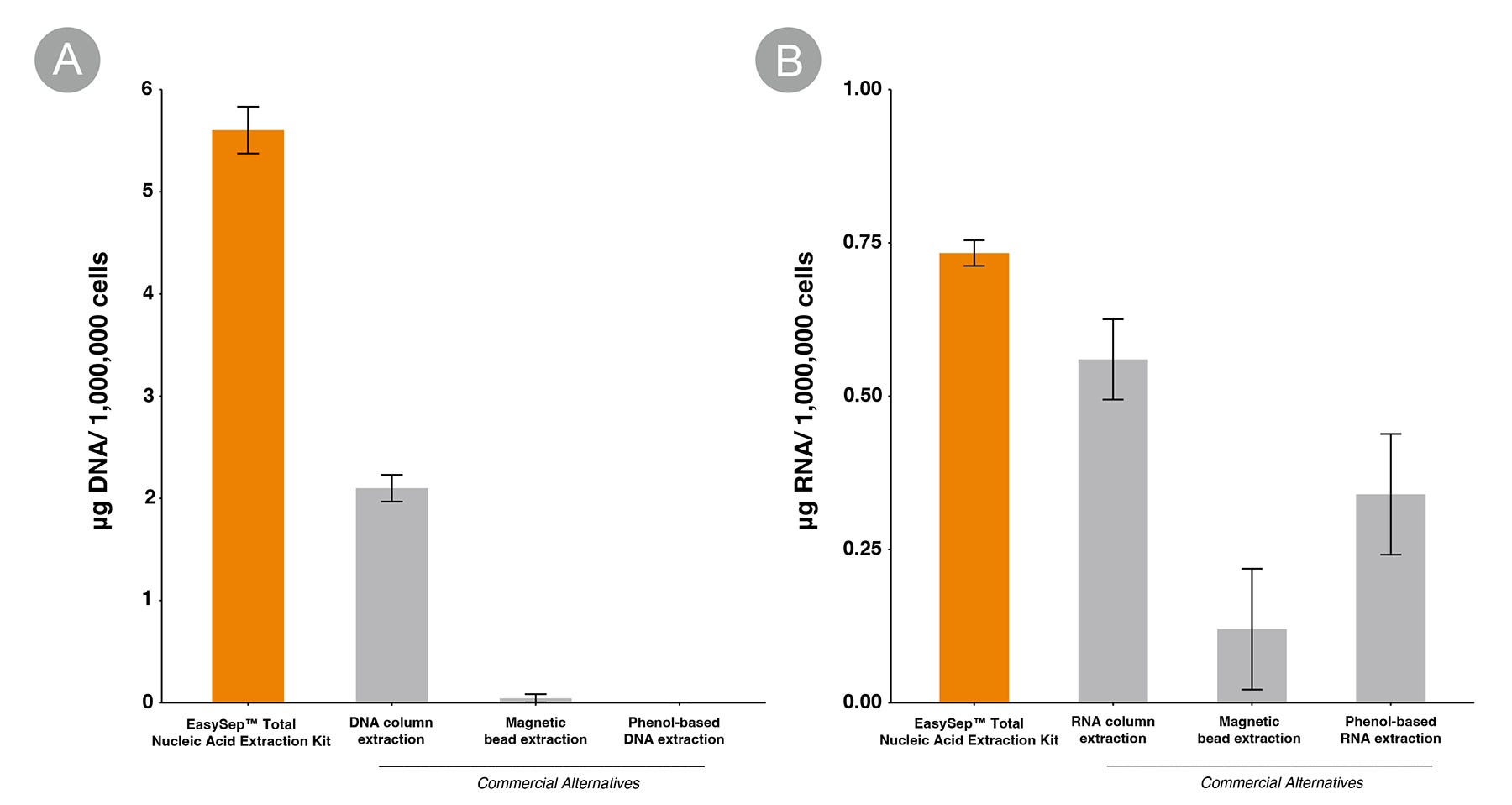
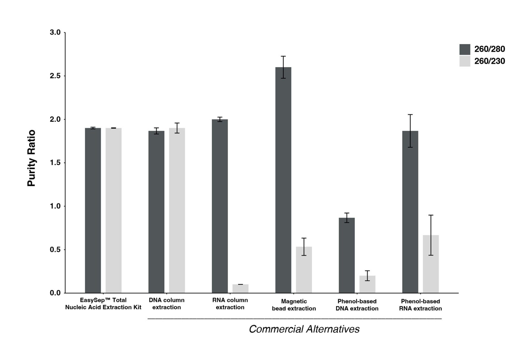
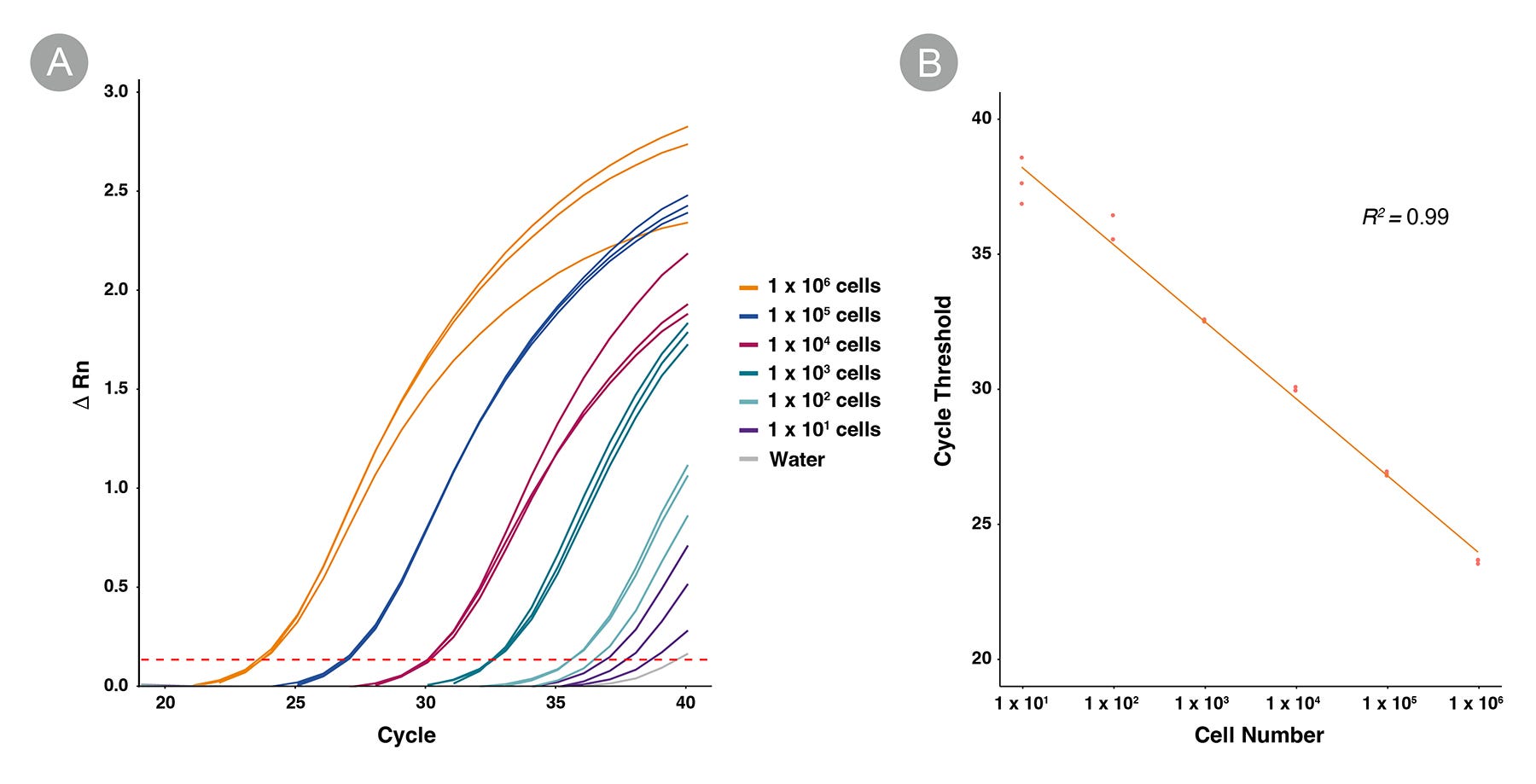
从全血、细胞悬液(如PBMCs、hPSCs、培养过的细胞)和EasySep™分选出的细胞中高效提取总核酸(DNA和RNA)。EasySep™总核酸提取试剂盒使用简单,可用于大通量提取,采用磁珠技术,可以避免使用离心柱或有毒试剂。该试剂盒提取的核酸纯度高,可直接用于下游应用,如qPCR。

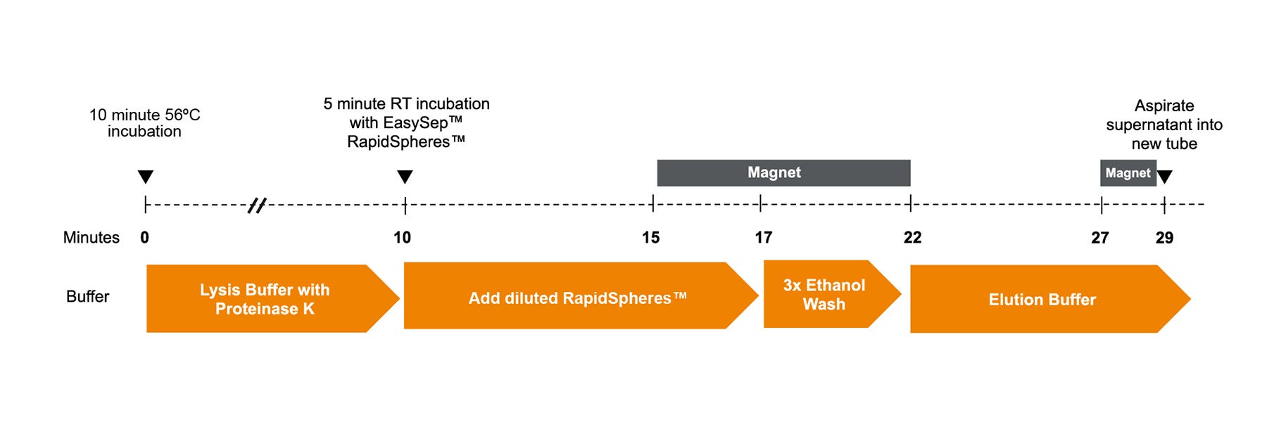
使用本试剂盒用EasySep™total nucleic Acid RapidSpheres™对样品中的总核酸进行磁性标记。然后,用磁铁(单独出售)处理样品——无需使用离心柱。使用移液管弃去未标记的非目标成分后,再从磁铁中取出样本,得到标记的核酸。可使用1.7mL微量离心管搭配ErythroClear™磁铁(产品号#01737);或为了扩大提取通量,使用96孔PCR微孔板搭配96孔PCR微孔板磁铁(产品号#100-1304)在96孔PCR板中处理样品。

磁体兼容性
ErythroClear™ Magnet (Catalog #01737) 96-Well PCR Microplate Magnet (Catalog #100-1304)
 
细胞类型
淋巴细胞,单个核细胞
 
应用
基因组编辑,核酸纯化
 
研究领域
癌症,免疫
 

Data Figures

Diagram of the EasySep™ Total Nucleic Acid Extraction Kit workflow.

Figure 1. EasySep™ Total Nucleic Acid Extraction Kit Workflow

Diagram of the standard extraction workflow. Time points at which samples must be placed in the magnet are indicated with gray boxes. EasySep™ Lysis Buffer and Proteinase K are added to the sample and incubated at 56°C for 10 minutes. Diluted EasySep™ Nucleic Acid RapidSpheres™ are added to the sample and incubated at room temperature (RT) for 5 minutes, then placed in the magnet for 2 minutes. While in the magnet, the sample is washed three times with a 70% ethanol wash solution, and the supernatant is removed. The pellet is resuspended with the elution buffer while removed from the magnet and incubated at room temperature for 5 minutes. The sample is then placed in the magnet for 2 minutes before the supernatant is aspirated to a new tube to obtain the extracted nucleic acids.

EasySep™ Total Nucleic Acid Extraction Kit shows improved DNA and RNA recovery relative to other commonly used methods.

Figure 2. EasySep™ Total Nucleic Acid Extraction Kit Shows Improved DNA and RNA Recovery Relative to Other Commonly Used Methods

Normalized recovery (μg per 1 x 10^6 cells) of (A) DNA and (B) RNA across EasySep™ Total Nucleic Acid Extraction Kit and other commonly used extraction methods. DNA and RNA concentrations were measured separately using the Qubit™ Fluorometer. Sample source: Leukopak, n = 3. Error bars represent ± 1 standard deviation.

EasySep™ Total Nucleic Acid Extraction Kit recovers nucleic acids of optimal purity when benchmarked against other commonly used extraction methods.

Figure 3. EasySep™ Total Nucleic Acid Extraction Kit Recovers Nucleic Acid of Optimal Purity

Purity ratios obtained using spectrophotometric absorbance measurements show that the nucleic acid recovered by the EasySep™ Total Nucleic Acid Extraction Kit is of optimal purity, benchmarked against other commonly used extraction methods. The 260/280 ratio, indicative of protein, phenol, and other contaminants within the extract that absorb at or near 280 nm, has an optimum purity ratio of ~ 1.8 for DNA and ~ 2.0 for RNA. The 260/230 ratio, a secondary measure of purity indicative of salt, phenol, and other organic compound contamination, has an optimal range of 1.8 - 2.2. Ratios that fall outside of these ranges are generally considered contaminated. Purity ratios were obtained using the Nanodrop™ Spectrophotometer. Sample source: Leukopak, n = 3. Error bars represent ± 1 standard deviation.

Results from a DNA-based qPCR assay with primers targeted to a non-genic region of chromosome 4 demonstrate that, DNA is detectable in extractions with a starting cell input of 10 cells and up to 1 x 10^6 cells, using the EasySep™ Total Nucleic Acid Extraction Kit.

Figure 4. EasySep™ Total Nucleic Acid Extraction Kit Is Capable of Extracting from As Few As 10 Cells

Results from a DNA-based qPCR assay using primers targeted to a non-genic region of chromosome 4 demonstrate that DNA is detectable in extractions with a starting cell input of 10 - 1 x 10^6 cells. (A) qPCR curves coloured by cell input. Dotted red line represents the cycle threshold (Ct; 0.139). Each individual curve represents 1 technical replicate, n = 3. (B) Inverse linear relationship between log(cell number input) and Ct value. Each data point represents 1 technical replicate, n = 3. Sample source: Leukopak.

Protocols and Documentation

Find supporting information and directions for use in the Product Information Sheet or explore additional protocols below.

Document Type
Product Name
Catalog #
Lot #
Language
Catalog #
100-1079
Lot #
All
Language
English
Catalog #
100-1079
Lot #
All
Language
English
Document Type
Safety Data Sheet 1
Catalog #
100-1079
Lot #
All
Language
English
Document Type
Safety Data Sheet 2
Catalog #
100-1079
Lot #
All
Language
English
Document Type
Safety Data Sheet 3
Catalog #
100-1079
Lot #
All
Language
English

Applications

This product is designed for use in the following research area(s) as part of the highlighted workflow stage(s). Explore these workflows to learn more about the other products we offer to support each research area.

Resources and Publications

Educational Materials (1)

Publications (1)

M1-like, but not M0- or M2-like, macrophages, reduce RSV infection of primary bronchial epithelial cells in a media-dependent fashion. N. J. Ronaghan et al. PloS one 2022

Abstract

Respiratory syncytial virus (RSV) is a common childhood infection that in young infants can progress into severe bronchiolitis and pneumonia. Disease pathogenesis results from both viral mediated and host immune processes of which alveolar macrophages play an important part. Here, we investigated the role of different types of alveolar macrophages on RSV infection using an in vitro co-culture model involving primary tissue-derived human bronchial epithelial cells (HBECs) and human blood monocyte-derived M0-like, M1-like, or M2-like macrophages. It was hypothesized that the in vitro model would recapitulate previous in vivo findings of a protective effect of macrophages against RSV infection. It was found that macrophages maintained their phenotype for the 72-hour co-culture time period and the bronchial epithelial cells were unaffected by the macrophage media. HBEC infection with RSV was decreased by M1-like macrophages but enhanced by M0- or M2-like macrophages. The medium used during the co-culture also impacted the outcome of the infection. This work demonstrates that alveolar macrophage phenotypes may have differential roles during epithelial RSV infection, and demonstrates that an in vitro co-culture model could be used to further investigate the roles of macrophages during bronchial viral infection.

更多信息

更多信息
Magnet Compatibility ErythroClear™ Magnet (Catalog #01737) 96-Well PCR Microplate Magnet (Catalog #100-1304)
PRODUCTS ARE FOR RESEARCH USE ONLY AND NOT INTENDED FOR HUMAN OR ANIMAL DIAGNOSTIC OR THERAPEUTIC USES UNLESS OTHERWISE STATED. FOR ADDITIONAL INFORMATION ON QUALITY AT STEMCELL, REFER TO WWW.STEMCELL.COM/COMPLIANCE.
版权 © 2025 STEMCELL Technologies 技术有限公司。保留所有权利。