产品号 #05888_C
成分明确的用于人ES和iPS细胞单细胞克隆生成的补充剂
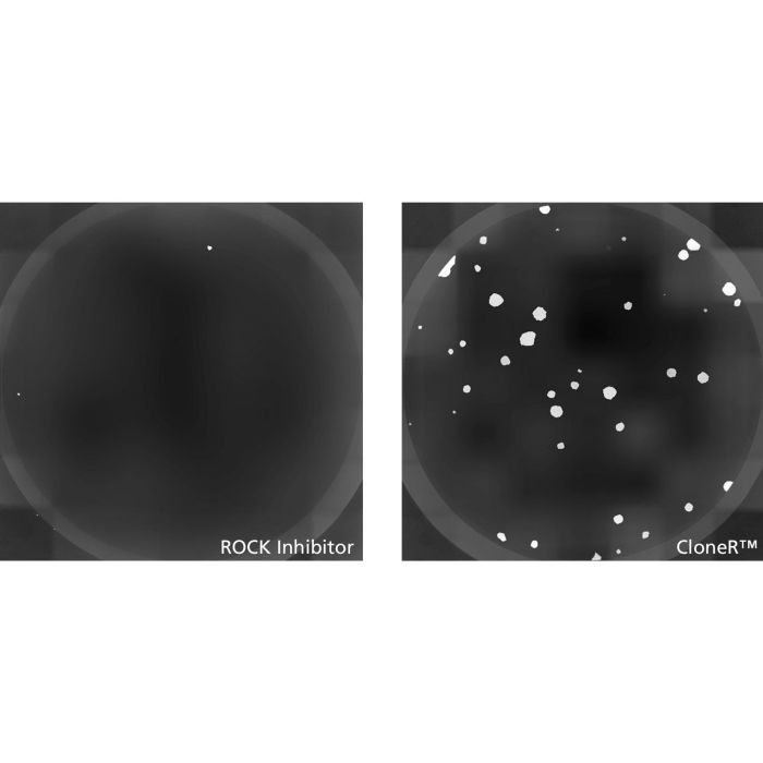
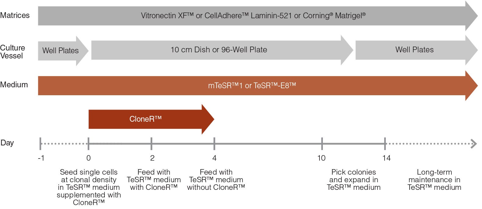
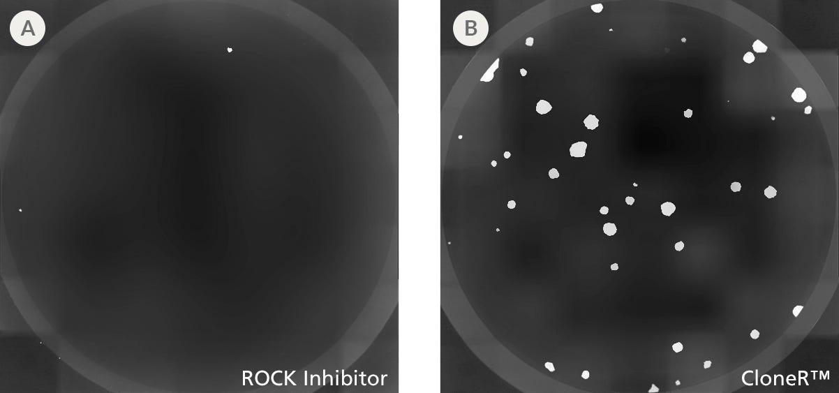
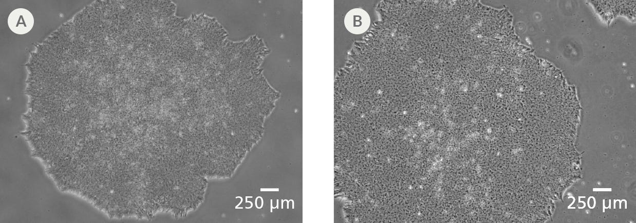
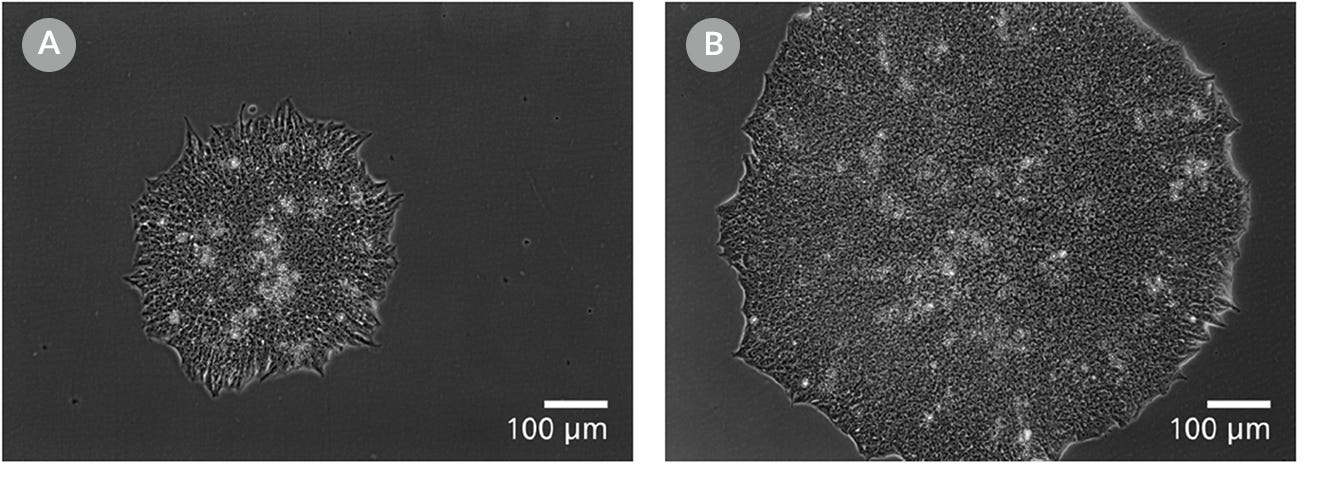
CloneR™ 是一种成分明确的无血清补充剂,旨在提高人胚胎干细胞 (ES 细胞) 和诱导性多能干细胞 (iPS 细胞) 的克隆生成效率和单细胞存活率。CloneR™ 可在无需进行单细胞适应的情况下,实现稳健的克隆细胞系生成,从而最大程度地减少获得遗传异常的风险。
CloneR™ 可与 TeSR™ 系列人ES和iPS细胞维持培养基以及您选择的细胞培养基质兼容使用。
如需在更多应用中改善单细胞存活率并提升克隆生成效率,可尝试我们的 CloneR™2 补充剂。
亚型
添加剂
细胞类型
多能干细胞
种属
人
应用
细胞培养
品牌
CloneR
研究领域
细胞系制备,疾病建模,干细胞生物学
制剂类别
无血清
Figure 1. hPSC Single-Cell Cloning Workflow with CloneR™
On day 0, human pluripotent stem cells (hPSCs) are seeded as single cells at clonal density (e.g. 25 cells/cm2) or sorted at 1 cell per well in 96-well plates in TeSR™ (mTeSR™1 or TeSR™-E8™) medium supplemented with CloneR™. On day 2, the cells are fed with TeSR™ medium containing CloneR™ supplement. From day 4, cells are maintained in TeSR™ medium without CloneR™. Colonies are ready to be picked between days 10 - 14. Clonal cell lines can be maintained long-term in TeSR™ medium.
Figure 2. CloneR™ Increases the Cloning Efficiency of hPSCs and is Compatible with Multiple hPSC Lines and Seeding Protocols
TeSR™ medium supplemented with CloneR™ increases hPSC cloning efficiency compared with cells plated in TeSR™ containing ROCK inhibitor. Cells were seeded (A) at clonal density (25 cells/cm2) in mTeSR™1 and TeSR™-E8™ and (B) by single-cell deposition using FACS (seeded at 1 cell/well) in mTeSR™1.
Figure 3. CloneR™ Increases the Cloning Efficiency of hPSCs at Low Seeding Densities
hPSCs plated in mTeSR™1 supplemented with CloneR™ demonstrated significantly increased cloning efficiencies compared to cells plated in mTeSR™1 containing ROCK inhibitor (10μM Y-27632). Shown are representative images of alkaline phosphatase-stained colonies at day 7 in individual wells of a 12-well plate. H1 human embryonic stem (hES) cells were seeded at clonal density (100 cells/well, 25 cells/cm2) in mTeSR™1 supplemented with (A) ROCK inhibitor or (B) CloneR™ on Vitronectin XF™ cell culture matrix.
Figure 4. CloneR™ Yields Larger Single-Cell Derived Colonies
hPSCs seeded in mTeSR™1 supplemented with CloneR™ result in larger colonies than cells seeded in mTeSR™1 containing ROCK inhibitor (10μM Y-27632). Shown are representative images of hPSC clones established after 7 days of culture in mTeSR™1 supplemented with (A) ROCK inhibitor or (B) CloneR™.
Figure 5. Clonal Cell Lines Established Using CloneR™ Display Characteristic hPSC Morphology
Clonal cell lines established using mTeSR™1 or TeSR™-E8™ medium supplemented with CloneR™ retain the prominent nucleoli and high nuclear-to-cytoplasmic ratio characteristic of hPSCs. Representative images at passage 7 after cloning are shown for clones derived from the parental (A) H1 hES cell and (B) WLS-1C human induced pluripotent stem (iPS) cell lines.
Figure 6. Clonal Cell Lines Established with CloneR™ Express High Levels of Undifferentiated Cell Markers
hPSC clonal cell lines established using mTeSR™1 supplemented with CloneR™ express comparable levels of undifferentiated cell markers, OCT4 (Catalog #60093) and TRA-1-60 (Catalog #60064), as the parental cell lines. (A) Clonal cell lines established from parental H1 hES cell line. (B) Clonal cell lines established from parental WLS-1C hiPS cell line. Data is presented between passages 5 - 7 after cloning and is shown as mean ± SEM; n = 2.
Figure 7. Clonal Cell Lines Established Using CloneR™ Display a Normal Karyotype
Representative karyograms of clones derived from parental (A) H1 hES cell and (B) WLS-1C hiPS cell lines demonstrate that the clonal lines established with CloneR™ have a normal karyotype. Cells were karyotyped 5 passages after cloning, with an overall passage number of 45 and 39, respectively.
Figure 8. Clonal Cell Lines Established Using CloneR™ Display Normal Growth Rates
Fold expansion of clonal cell lines display similar growth rates to parental cell lines. Shown are clones (red) and parental cell lines (gray) for (A) H1 hES cell and (B) WLS-1C hiPS cell lines.
Figure 9. Representative Cell Morphology 24 Hours After RNP Electroporation in mTeSR™1 and mTeSR™ Plus
H1-eGFP ES cells were plated in (A) mTeSR™1 and (B) mTeSR™ Plus and supplemented with CloneR™ immediately following RNP electroporation. Images were taken 24 hours after electroporation.
Figure 10. Clones Derived in mTeSR™ Plus are Larger and Ready to Be Picked at an Earlier Timepoint
Representative images of human ES (H9) colonies taken 8 days following singlecell plating at clonal density (25 cells/cm²) in either (A) mTeSR™1 or (B) mTeSR™ Plus supplemented with CloneR™ on CellAdhere™ Vitronectin™ XF™-coated plates.
Find supporting information and directions for use in the Product Information Sheet or explore additional protocols below.
This product is designed for use in the following research area(s) as part of the highlighted workflow stage(s). Explore these workflows to learn more about the other products we offer to support each research area.
Thank you for your interest in IntestiCult™ Organoid Growth Medium (Human). Please provide us with your contact information and your local representative will contact you with a customized quote. Where appropriate, they can also assist you with a(n):
Estimated delivery time for your area
Product sample or exclusive offer
In-lab demonstration
Species | Human |
---|---|
Formulation Category | Serum-Free |
细胞分离溶液
用于提高人胚胎干细胞和有道多能干细胞在单细胞工作流程中的存活率添加物
与 TeSR™ 维持培养基配合使用,用于维持人胚胎干细胞(ES)和诱导多能干细胞(iPS)的基质
cGMP标准、无饲养层的人胚胎干细胞(ES)和诱导多能干细胞(iPS)维持培养基
扫描二维码或搜索微信号STEMCELLTech,即可关注我们的微信平台,第一时间接收丰富的技术资源和最新的活动信息。
如您有任何问题,欢迎发消息给STEMCELLTech微信公众平台,或与我们通过电话/邮件联系:400 885 9050 INFO.CN@STEMCELL.COM。