抗小鼠SSEA-1(CD15)抗体,克隆MC-480

抗人、小鼠、大鼠SSEA-1(CD15)的小鼠单克隆IgM抗体

产品号 #(选择产品)

产品号 #60060_C

抗人、小鼠、大鼠SSEA-1(CD15)的小鼠单克隆IgM抗体

总览

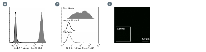
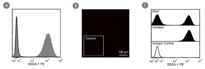
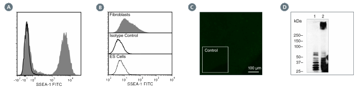
MC-480抗体与末端碳水化合物表位,阶段特异性胚胎抗原-1 (SSEA-1)反应,SSEA-1表达于小鼠早期胚胎,小鼠胚胎癌(EC),胚胎干细胞(ES)和小鼠及人胚胎生殖(EG)细胞表面的一种大分子量(> 200 kDa)糖蛋白上。SSEA-1在未分化的人EC、ES或诱导多能干细胞(iPS)或恒河猴ES细胞系上不表达。其在小鼠胚胎干细胞中的表达在分化过程中降低,而在人ES细胞中,其表达在分化过程中会上调。SSEA-1也存在于成人粒细胞和单核细胞中,标记为CD15, MC-480抗体识别这些细胞类型上的CD15标记。据报道,SSEA-1在细胞粘附和迁移以及细胞分化调控中发挥作用。

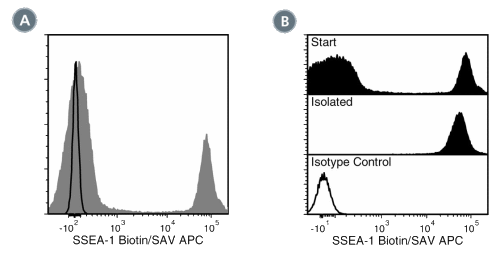
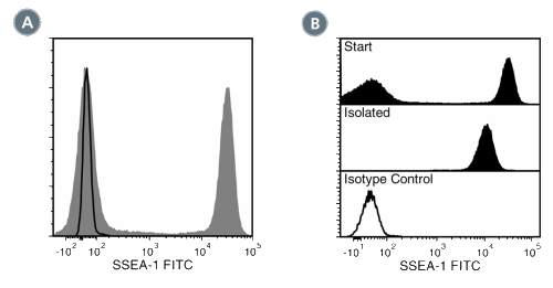
该抗体克隆已通过EasySep™试剂盒分离的细胞纯度评估验证,包括EasySep™HLA全血CD15正选试剂盒(产品号#18681HLA;可观察到部分阻断),并用于标记在TeSR™-E8™(产品号#05940),mTeSR™1(产品号#85850)和TeSR™2(产品号#05860)中生长的人 ES和iPS细胞。

亚型
一抗
 
靶抗原
SSEA-1 (CD15)
 
别名
3-FAL,CD15,Lewis X,SSEA1,阶段特异性胚胎抗原1,X-半抗原
 
活性物种
人,小鼠,大鼠
 
偶联
Alexa Fluor 488,Biotin 或 生物素,FITC,PE,未偶联的
 
宿主物种
小鼠
 
细胞类型
多能干细胞
 
种属
人,小鼠,大鼠
 
应用
细胞分选,流式细胞术,免疫细胞化学,免疫荧光,免疫组化,免疫沉淀,Western印迹
 
研究领域
干细胞生物学
 
克隆
MC-480
 
基因编号
14345
 
同种型
IgM,kappa
 

Data Figures

Data for Alexa Fluor® 488-Conjugated

Figure 1. Data for Alexa Fluor® 488-Conjugated

(A) Flow cytometry analysis of human whole blood nucleated cells labeled with Anti-Human SSEA-1 (CD15) Antibody, Clone MC-480, Alexa Fluor® 488 (filled histogram) or Mouse IgM, kappa Isotype Control Antibody, Clone MM-30, Alexa Fluor® 488 (Catalog #60069AD) (solid line histogram). SSEA-1 is highly expressed on granulocytes.
(B) Flow cytometry analysis of human HT1080 fibrosarcoma cells labeled with Anti-Human SSEA-1 (CD15) Antibody, Clone MC-480, Alexa Fluor® 488 (filled histogram). Labeling of human HT1080 fibrosarcoma (solid line histogram) or ES cells (negative control; dashed line histogram) with a mouse IgM, kappa isotype control antibody (Anti-Human TRA-1-60 Antibody, Clone TRA-1-60R, Alexa Fluor® 488; Catalog #60064AD) is shown. SSEA-1 is not expressed on undifferentiated human ES cells.
(C) Human ES cells were cultured in mTeSR™1 on Corning® Matrigel®-coated glass slides, then fixed and labeled with Anti-Human SSEA-1 (CD15) Antibody, Clone MC-480, Alexa Fluor® 488. Inset shows cells labeled with Mouse IgM, kappa Isotype Control Antibody, Clone MM-30, Alexa Fluor® 488.

Data for Biotin-Conjugated

Figure 2. Data for Biotin-Conjugated

(A) Flow cytometry analysis of human buffy coat nucleated cells labeled with Anti-Human SSEA-1 Antibody, Clone MC-480, Biotin, followed by streptavidin (SAV) APC (filled histogram), or a mouse IgM, kappa biotin isotype control antibody, followed by SAV APC (solid line histogram). (B) Flow cytometry analysis of human buffy coat nucleated cells processed with the EasySep™ HLA Whole Blood CD15 Positive Selection Kit and labeled with Anti-Human SSEA-1 Antibody, Clone MC-480, Biotin, followed by SAV APC. Histograms show labeling of buffy coat nucleated cells (Start) and isolated cells (Isolated). Labeling with a mouse IgM, kappa biotin isotype control antibody, followed by SAV APC is shown (solid line histogram).

Data for FITC-Conjugated

Figure 3. Data for FITC-Conjugated

(A) Flow cytometry analysis of human buffy coat nucleated cells labeled with Anti-Human SSEA-1 Antibody, Clone MC-480, FITC (filled histogram) or a mouse IgM, kappa FITC Isotype control antibody (solid line histogram). SSEA-1 (CD15) is highly expressed on granulocytes. (B) Flow cytometry analysis of human buffy coat nucleated cells processed with the EasySep™ HLA CD15 WB Positive Selection Kit and labeled with Anti-Human SSEA-1 Antibody, Clone MC-480, FITC. Histograms show labeling of buffy coat nucleated cells (Start) and isolated cells (Isolated). Labeling of start cells with a mouse IgM, kappa FITC Isotype control antibody is shown (solid line histogram).

Data for PE-Conjugated

Figure 4. Data for PE-Conjugated

(A) Flow cytometry analysis of human whole blood nucleated cells labeled with Anti-Human SSEA-1 (CD15) Antibody, Clone MC-480, PE (filled histogram) or Mouse IgM, kappa Isotype Control Antibody, Clone MM-30, PE (Catalog #60069PE) (solid line histogram). SSEA-1 is highly expressed on granulocytes.
(B) Human ES cells were cultured in mTeSR™1 on Corning® Matrigel®-coated glass slides, then fixed and labeled with Anti-Human SSEA-1 (CD15) Antibody, Clone MC-480, PE. Inset shows cells labeled with Mouse IgM, kappa Isotype Control Antibody, Clone MM-30, PE. SSEA-1 is not expressed on undifferentiated human ES cells.
(C) Flow cytometry analysis of human buffy coat nucleated cells processed with the EasySep™ HLA CD15 WB Positive Selection Kit and labeled with Anti-Human SSEA-1 (CD15) Antibody, Clone MC-480, PE. Histograms show labeling of buffy coat nucleated cells (Start) and isolated cells (Isolated). Labeling with Mouse IgM, kappa Isotype Control Antibody, Clone MM-30, PE is shown (solid line histogram).

Data for Unconjugated

Figure 5. Data for Unconjugated

(A) Flow cytometry analysis of human whole blood nucleated cells labeled with Anti-Human SSEA-1 (CD15) Antibody, Clone MC-480, followed by Goat Anti-Mouse IgG (H+L) Antibody, Polyclonal, FITC (Catalog #60138FI) (filled histogram), or Mouse IgM, kappa Isotype Control Antibody, Clone MM-30 (Catalog #60069), followed by Goat Anti-Mouse IgG (H+L) Antibody, Polyclonal, FITC (solid line histogram). SSEA-1 is highly expressed on granulocytes.
(B) Flow cytometry analysis of human HT1080 fibrosarcoma cells labeled with Anti-Human SSEA-1 (CD15) Antibody, Clone MC-480, followed by goat antimouse IgG, FITC (filled histogram). Labeling of human HT1080 fibrosarcoma cells (solid line histogram) or H1 ES cells (negative control; dashed line histogram) with a mouse IgM, kappa isotype control antibody (Anti-Human TRA-1-60 Antibody, Clone TRA-1-60R; Catalog #60064) is shown.
(C) Human ES cells were cultured in mTeSR™1 on Corning® Matrigel®-coated glass slides, then fixed and labeled with Anti-Human SSEA-1 (CD15) Antibody, Clone MC-480, followed by goat anti-mouse IgG, FITC. Inset shows cells labeled with Mouse IgM, kappa Isotype Control Antibody, Clone MM- 30, followed by goat anti-mouse IgG, FITC. SSEA-1 is not expressed on undifferentiated human ES cells.
(D) Western blot analysis of denatured/reduced cell lysates from human ES cells (negative control; lane 1) or HT1080 fibrosarcoma cells (lane 2) with Anti-Human SSEA-1 (CD15) Antibody, Clone MC-480.

Protocols and Documentation

Find supporting information and directions for use in the Product Information Sheet or explore additional protocols below.

Document Type
Product Name
Catalog #
Lot #
Language
Catalog #
60060FI, 60060FI.1
Lot #
All
Language
English
Catalog #
60060AD, 60060AD.1
Lot #
All
Language
English
Catalog #
60060
Lot #
All
Language
English
Catalog #
60060BT
Lot #
All
Language
English
Catalog #
60060PE.1, 60060PE
Lot #
All
Language
English
Document Type
Safety Data Sheet
Catalog #
60060FI, 60060FI.1
Lot #
All
Language
English
Document Type
Safety Data Sheet
Catalog #
60060AD, 60060AD.1
Lot #
All
Language
English
Document Type
Safety Data Sheet
Catalog #
60060
Lot #
All
Language
English
Document Type
Safety Data Sheet
Catalog #
60060BT
Lot #
All
Language
English
Document Type
Safety Data Sheet
Catalog #
60060PE.1, 60060PE
Lot #
All
Language
English

Applications

This product is designed for use in the following research area(s) as part of the highlighted workflow stage(s). Explore these workflows to learn more about the other products we offer to support each research area.

Resources and Publications

Educational Materials (3)

Publications (1)

OCT4 as a target of miR-34a stimulates p63 but inhibits p53 to promote human cell transformation Ng WL et al. Cell death & disease 2014 JAN

Abstract

Human cell transformation is a key step for oncogenic development, which involves multiple pathways; however, the mechanism remains unclear. To test our hypothesis whether cell oncogenic transformation shares some mechanisms with the process of reprogramming non-stem cells to induced pluripotent stem cells (iPSC), we studied the relationship among the key factors for promoting or inhibiting iPSC in radiation-transformed human epithelial cell lines derived from different tissues (lung, breast and colon). We unexpectedly found that p63 and OCT4 were highly expressed (accompanied by low expressed p53 and miR-34a) in all transformed cell lines examined when compared with their non-transformed counterparts. We further elucidated the relationship of these factors: the 3p strand of miR-34a directly targeted OCT4 by binding to the 3′ untranslated region (3′-UTR) of OCT4 and, OCT4, in turn, stimulated p63 but inhibited p53 expression by binding to a specific region of the p63 or p53 promoter. Moreover, we revealed that the effects of OCT4 on promoting cell oncogenic transformation were by affecting p63 and p53. These results support that a positive loop exists in human cells: OCT4 upregulation as a consequence of inhibition of miR-34a, promotes p63 but suppresses p53 expression, which further stimulates OCT4 upregulation by downregulating miR-34a. This functional loop contributes significantly to cell transformation and, most likely, also to the iPSC process.

更多信息

更多信息
Species Human, Mouse, Rat
Clone MC-480
Gene Id 14345
Alternative Names 3-FAL, CD15, Lewis X, SSEA1, Stage-specific embryonic antigen 1, X-hapten
Isotype IgM, kappa
Legal Statement: Alexa Fluor is a registered trademark of Life Technologies Corporation. Antibodies conjugated to Alexa Fluor® are licensed for internal research use only and sale is expressly conditioned on the buyer not using the antibody for manufacturing, performing a service or medical test, or otherwise generating revenue. For use other than research, contact Life Technologies Corporation, 5791 Van Allen Way, Carlsbad, CA 92008 USA or outlicensing@lifetech.com. PRODUCTS ARE FOR RESEARCH USE ONLY AND NOT INTENDED FOR HUMAN OR ANIMAL DIAGNOSTIC OR THERAPEUTIC USES UNLESS OTHERWISE STATED. FOR ADDITIONAL INFORMATION ON QUALITY AT STEMCELL, REFER TO WWW.STEMCELL.COM/COMPLIANCE.
版权 © 2025 STEMCELL Technologies 技术有限公司。保留所有权利。