产品号 #60036_C
抗小鼠CD150(SLAM)大鼠单克隆IgG2a抗体
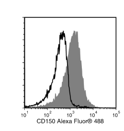
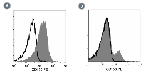
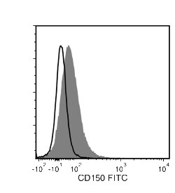
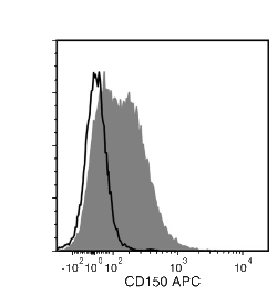
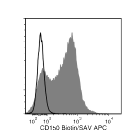
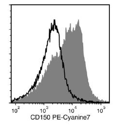
TC15-12F12.2抗体与CD150(信号淋巴细胞活化分子,SLAM)发生反应。CD150是一种分子量约为75 kDa的I型跨膜糖蛋白,作为细胞粘附分子和/或辅助受体,在免疫反应中发挥多种作用。它在T细胞、未成熟胸腺细胞、B细胞、树突状细胞、巨噬细胞和内皮细胞中存在差异表达。其表达模式因细胞类型和活化状态而异。T细胞、B细胞和树突状细胞活化后,CD150会快速诱导表达,且T细胞的合成主要在Th1细胞克隆上进行,而非Th2细胞克隆。CD150介导的TCR激活T细胞的共刺激作用可增强Th1细胞产生IFN-γ,而这种应答可通过TC15-12F12.2抗体的结合得到增强。 CD150被认为通过与细胞内蛋白酪氨酸磷酸酶SHP-2结合来介导信号转导。CD150还在造血细胞发育中发挥作用,并且是检测多能造血干细胞的有效标记物(与CD48和CD41等其他标记物协同作用)。CD150在非多能造血祖细胞上不表达。
亚型
一抗
靶抗原
CD150 (SLAM)
别名
IPO-3,信号淋巴细胞活化分子
活性物种
小鼠
偶联
Alexa Fluor 488,APC,Biotin 或 生物素,PE,PE-Cyanine7,未偶联的
宿主物种
Hamster,大鼠
细胞类型
造血干/祖细胞
种属
小鼠
应用
流式细胞术,功能学筛选,免疫荧光,免疫组化,免疫沉淀
研究领域
干细胞生物学
克隆
TC15-12F12.2
基因编号
27218
同种型
IgG2a,lambda
Figure 1. Data for Alexa Fluor® 488-Conjugated
Flow cytometry analysis of C57BL/6 mouse splenocytes labeled with Anti-Mouse CD150 Antibody, Clone TC15-12F12.2, Alexa Fluor® 488 (filled histogram) or a rat IgG2a, lambda Alexa Fluor® 488 isotype control antibody (solid line histogram).
Figure 2. Data for PE-Conjugated
(A) Flow cytometry analysis of C57BL/6 mouse splenocytes labeled with Anti-Mouse CD150 Antibody, Clone TC15-12F12.2, PE (filled histogram) or a rat IgG2a, lambda PE isotype control antibody (solid line histogram). (B) Flow cytometry analysis of C57BL/6 mouse bone marrow cells labeled with Anti-Mouse CD150 Antibody, Clone TC15-12F12.2, PE (filled histogram) or a rat IgG2a, lambda PE isotype control antibody (solid line histogram).
Figure 3. Data for Unconjugated
Flow cytometry analysis of C57BL/6 mouse splenocytes labeled with Anti-Mouse CD150 Antibody, Clone TC15-12F12.2, followed by a mouse anti-rat IgG2a antibody, FITC (filled histogram), or a rat IgG2a isotype control antibody followed by a mouse anti-rat IgG2a antibody, FITC (solid line histogram).
Figure 4. Data for APC-Conjugated
Flow cytometry analysis of C57BL/6 mouse splenocytes labeled with Anti-Mouse CD150 Antibody, Clone TC15-12F12.2, APC (filled histogram) or a rat IgG2a isotype control antibody, APC (solid line histogram).
Figure 5. Data for Biotin-Conjugated
Flow cytometry analysis of C57BL/6 mouse splenocytes labeled with Anti-Mouse CD150 Antibody, Clone TC15-12F12.2, Biotin followed by streptavidin (SAV) APC (filled histogram), or a biotinylated rat IgG2a isotype control antibody followed by SAV APC (solid line histogram).
(A) Primary human airway epithelial cells were cultured in PneumaCult™-ALI Medium (Catalog #05001) at the air-liquid interface, then cryo-sectioned and labeled with Anti-Human MUC1 (CD227) Antibody, Clone 16A, followed by a goat anti-rabbit IgG antibody, Alexa Fluor® 594 (red), and an anti-human NGF Receptor/p75NTR (CD271) antibody, followed by a donkey anti-mouse IgG antibody, Alexa Fluor® 488 (green). (B) Flow cytometry analysis of human peripheral blood lymphocytes following stimulation with PHA for 3 days. Cells were labeled with Anti-Human MUC1 (CD227) Antibody, Clone 16A, followed by an anti-mouse IgG1 antibody, PE and anti-human CD3 antibody, Clone HIT3a, Pacific Blue™. (C) Flow cytometry analysis of human peripheral blood lymphocytes following stimulation with PHA for 3 days. Cells were labeled with Mouse IgG1, kappa Isotype Control Antibody, Clone MOPC-21 (Catalog #60070), followed by an anti-mouse IgG1 antibody, PE, and anti-human CD3 antibody, clone HIT3a, Pacific Blue™.
(A) Flow cytometry analysis of human airway epithelial cells cultured in PneumaCult™-ALI Medium (Catalog #05001) at the air-liquid interface. Cells were enzymatically dissociated and labeled with Anti-Human MUC1 (CD227) Antibody, Clone 16A, PE (filled histogram) or Mouse IgG1, kappa Isotype Control Antibody, Clone MOPC-21, PE (Catalog #60070PE, solid line histogram). (C) Flow cytometry analysis human peripheral blood lymphocytes following stimulation with PHA for 3 days. Cells were labeled with Anti-Human MUC1 (CD227) Antibody, Clone 16A, PE and anti-human CD3 antibody, clone HIT3a, Pacific Blue™. (D) Flow cytometry analysis of PHA-activated human peripheral blood lymphocytes following stimulation with PHA for 3 days. Cells were labeled with Mouse IgG1, kappa Isotype Control Antibody, Clone MOPC-21, PE, and anti-human CD3 antibody, clone HIT3a, Pacific Blue™.
Flow cytometry analysis of C57BL/6 mouse splenocytes labeled with Anti-Mouse CD150 Antibody, Clone TC15-12F12.2, PE-Cyanine7 (filled histogram) or a rat IgG2a isotype control antibody, PE-Cyanine7 (solid line histogram).
Find supporting information and directions for use in the Product Information Sheet or explore additional protocols below.
This product is designed for use in the following research area(s) as part of the highlighted workflow stage(s). Explore these workflows to learn more about the other products we offer to support each research area.
Thank you for your interest in IntestiCult™ Organoid Growth Medium (Human). Please provide us with your contact information and your local representative will contact you with a customized quote. Where appropriate, they can also assist you with a(n):
Estimated delivery time for your area
Product sample or exclusive offer
In-lab demonstration
Species | Mouse |
---|---|
Clone | TC15-12F12.2 |
Gene Id | 27218 |
Alternative Names | IPO-3, Signaling lymphocyte activation molecule |
Isotype | IgG2a, lambda |
含有重组细胞因子(包括EPO)的甲基纤维素基培养基用于小鼠细胞
用于培养和扩增造血细胞的无血清培养基
抗小鼠EPCR(CD201)大鼠单克隆IgG2b抗体
大鼠单克隆IgG2a, kappa同型对照抗体
扫描二维码或搜索微信号STEMCELLTech,即可关注我们的微信平台,第一时间接收丰富的技术资源和最新的活动信息。
如您有任何问题,欢迎发消息给STEMCELLTech微信公众平台,或与我们通过电话/邮件联系:400 885 9050 INFO.CN@STEMCELL.COM。