抗人CD32抗体,克隆IV.3

小鼠单克隆IgG2b抗体,抗人CD32

产品号 #(选择产品)

产品号 #60012_C

小鼠单克隆IgG2b抗体,抗人CD32

总览

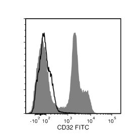
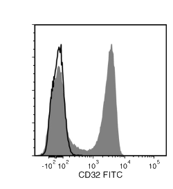
IV.3抗体识别人CD32 (FcγRII),CD32是一种~40 kDa的1型跨膜糖蛋白,介导多种功能,包括吞噬、细胞毒性、免疫调节和血小板聚集。CD32由3个基因(A、B、C)编码,通过mRNA的选择性剪接产生至少6个异构体,即IIa1、IIa2、IIb1、IIb2、IIb3和IIc。单核/巨噬细胞、胎盘滋养细胞和内皮细胞表达所有亚型。此外,B细胞表达IIb亚型;血小板、粒细胞表达IIa亚型,B细胞弱表达IIa亚型。NK细胞和中性粒细胞表达IIc亚型。CD32与IgG单体的Fc段结合较弱,但与IgG聚集体和免疫复合物结合较强。这些相互作用可导致基于抗体的检测和细胞分选实验中的非特异性标记,而IV.3抗体可作为阻断抗体来减少非特异性结合。IV.3抗体与CD32的IIa亚型结合最强,其表位位于配体结合位点的结构域2中的132 - 137 氨基酸[FSHLDP]。流式细胞术分析显示,克隆FLI8.26可以阻断IV.3抗体的结合,表明这两个克隆可能具有共同或重叠的表位。

该抗体克隆已被验证可用于评估EasySep™试剂盒(包括EasySep™人T细胞富集试剂盒(产品号 #19051)和EasySep™人CD4+ T细胞富集试剂盒(产品号#19052))分选的细胞纯度。

亚型
一抗
 
靶抗原
CD32
 
别名
FCR II,FcγRII
 
活性物种

 
偶联
FITC,未偶联的
 
宿主物种
小鼠
 
细胞类型
B 细胞,粒细胞及其亚群,单核细胞
 
种属

 
应用
细胞分选,流式细胞术,功能学筛选,免疫细胞化学,免疫组化,中和及阻断,Western印迹
 
研究领域
免疫
 
克隆
IV.3
 
基因编号
2212
 
同种型
IgG2b,kappa
 

Data Figures

Data for Unconjugated

Figure 1. Data for Unconjugated

Flow cytometry analysis of human peripheral blood mononuclear cells (PBMCs) labeled with Anti-Human CD32 Antibody, Clone IV.3, followed by Goat Anti-Mouse IgG (H+L) Antibody, Polyclonal, FITC (Catalog #60138FI; filled histogram), or Mouse IgG2b, kappa Isotype Control Antibody, Clone MPC-11 (Catalog #60072), followed by Goat Anti-Mouse IgG (H+L) Antibody, Polyclonal, FITC (solid line histogram).

Data for FITC-Conjugated

Figure 2. Data for FITC-Conjugated

Flow cytometry analysis of human peripheral blood mononuclear cells (PBMCs) labeled with Anti-Human CD32 Antibody, Clone IV.3, FITC (filled histogram) or Mouse IgG2b, kappa Isotype Control Antibody, Clone MPC-11, FITC (Catalog #60072FI) (solid line histogram).

Protocols and Documentation

Find supporting information and directions for use in the Product Information Sheet or explore additional protocols below.

Document Type
Product Name
Catalog #
Lot #
Language
Catalog #
60012FI.1
Lot #
All
Language
English
Catalog #
60012
Lot #
All
Language
English
Catalog #
100-1574
Lot #
All
Language
English
Document Type
Safety Data Sheet
Catalog #
60012FI.1, 100-1574
Lot #
All
Language
English
Document Type
Safety Data Sheet
Catalog #
60012
Lot #
All
Language
English

Applications

This product is designed for use in the following research area(s) as part of the highlighted workflow stage(s). Explore these workflows to learn more about the other products we offer to support each research area.

Resources and Publications

Educational Materials (2)

Publications (2)

Transmembrane Pickets Connect Cyto- and Pericellular Skeletons Forming Barriers to Receptor Engagement. Freeman SA et al. Cell 2018 JAN

Abstract

Phagocytic receptors must diffuse laterally to become activated upon clustering by multivalent targets. Receptor diffusion, however, can be obstructed by transmembrane proteins (pickets") that are immobilized by interacting with the cortical cytoskeleton. The molecular identity of these pickets and their role in phagocytosis have not been defined. We used single-molecule tracking to study the interaction between Fcγ receptors and CD44 an abundant transmembrane protein capable of indirect association with F-actin hence likely to serve as a picket. CD44 tethers reversibly to formin-induced actin filaments curtailing receptor diffusion. Such linear filaments predominate in the trailing end of polarized macrophages where receptor mobility was minimal. Conversely receptors were most mobile at the leading edge where Arp2/3-driven actin branching predominates. CD44 binds hyaluronan anchoring a pericellular coat that also limits receptor displacement and obstructs access to phagocytic targets. Force must be applied to traverse the pericellular barrier enabling receptors to engage their targets.
Mast cells form antibody-dependent degranulatory synapse for dedicated secretion and defence. Joulia R et al. Nature communications 2015 JAN

Abstract

Mast cells are tissue-resident immune cells that play a key role in inflammation and allergy. Here we show that interaction of mast cells with antibody-targeted cells induces the polarized exocytosis of their granules resulting in a sustained exposure of effector enzymes, such as tryptase and chymase, at the cell-cell contact site. This previously unidentified mast cell effector mechanism, which we name the antibody-dependent degranulatory synapse (ADDS), is triggered by both IgE- and IgG-targeted cells. ADDSs take place within an area of cortical actin cytoskeleton clearance in the absence of microtubule organizing centre and Golgi apparatus repositioning towards the stimulating cell. Remarkably, IgG-mediated degranulatory synapses also occur upon contact with opsonized Toxoplasma gondii tachyzoites resulting in tryptase-dependent parasite death. Our results broaden current views of mast cell degranulation by revealing that human mast cells form degranulatory synapses with antibody-targeted cells and pathogens for dedicated secretion and defence.

更多信息

更多信息
Species Human
Clone IV.3
Gene Id 2212
Alternative Names FCR II, FcγRII
Isotype IgG2b, kappa
PRODUCTS ARE FOR RESEARCH USE ONLY AND NOT INTENDED FOR HUMAN OR ANIMAL DIAGNOSTIC OR THERAPEUTIC USES UNLESS OTHERWISE STATED. FOR ADDITIONAL INFORMATION ON QUALITY AT STEMCELL, REFER TO WWW.STEMCELL.COM/COMPLIANCE.
版权 © 2025 STEMCELL Technologies 技术有限公司。保留所有权利。