


产品号 #60155_C
Mouse monoclonal IgG1 antibody against human MUC1 (CD227)
The 16A antibody reacts with human MUC1, a large (> 250 kDa) heavily glycosylated type 1 transmembrane protein expressed on the surface of most glandular and ductal epithelial cells and a variety of hematopoietic cells. A characteristic feature of the MUC1 glycoprotein is a core domain composed of a variable number of tandem repeats and multiple oligosaccharide side chains. Because the extracellular portion of MUC1 can extend beyond most cell surface proteins, it is thought to play a role in cell-cell and cell-substrate adhesion. The protein is highly expressed by a majority of human adenocarcinomas and is associated with a poor prognosis. In the mammary gland, MUC1 is localized on the apical plasma membrane of luminal epithelial cells. The clone 16A antibody has a higher affinity for the glycosylated form of MUC1.
Subtype
Primary Antibodies
Target Antigen
MUC1 (CD227)
Alternative Names
CD227, EMA, Episialin, Epithelial membrane antigen, HMFG antigen, MAM6, Mucin 1, PEM, Polymorphic epithelial mucin
Reactive Species
Human
Conjugation
APC, PE, Unconjugated
Host Species
Mouse
Cell Type
Airway Cells
Application
Flow Cytometry, Immunocytochemistry, Immunofluorescence, Immunohistochemistry
Area of Interest
Epithelial Cell Biology
Clone
16A
Gene ID
4582
Isotype
IgG1, lambda
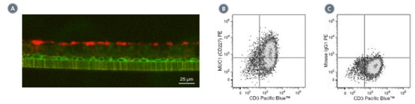
(A) Primary human airway epithelial cells were cultured in PneumaCult™-ALI Medium (Catalog #05001) at the air-liquid interface, then cryo-sectioned and labeled with Anti-Human MUC1 (CD227) Antibody, Clone 16A, followed by a goat anti-rabbit IgG antibody, Alexa Fluor® 594 (red), and an anti-human NGF Receptor/p75NTR (CD271) antibody, followed by a donkey anti-mouse IgG antibody, Alexa Fluor® 488 (green). (B) Flow cytometry analysis of human peripheral blood lymphocytes following stimulation with PHA for 3 days. Cells were labeled with Anti-Human MUC1 (CD227) Antibody, Clone 16A, followed by an anti-mouse IgG1 antibody, PE and anti-human CD3 antibody, Clone HIT3a, Pacific Blue™. (C) Flow cytometry analysis of human peripheral blood lymphocytes following stimulation with PHA for 3 days. Cells were labeled with Mouse IgG1, kappa Isotype Control Antibody, Clone MOPC-21 (Catalog #60070), followed by an anti-mouse IgG1 antibody, PE, and anti-human CD3 antibody, clone HIT3a, Pacific Blue™.
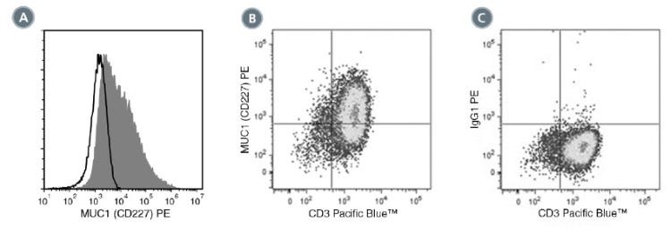
(A) Flow cytometry analysis of human airway epithelial cells cultured in PneumaCult™-ALI Medium (Catalog #05001) at the air-liquid interface. Cells were enzymatically dissociated and labeled with Anti-Human MUC1 (CD227) Antibody, Clone 16A, PE (filled histogram) or Mouse IgG1, kappa Isotype Control Antibody, Clone MOPC-21, PE (Catalog #60070PE, solid line histogram). (C) Flow cytometry analysis human peripheral blood lymphocytes following stimulation with PHA for 3 days. Cells were labeled with Anti-Human MUC1 (CD227) Antibody, Clone 16A, PE and anti-human CD3 antibody, clone HIT3a, Pacific Blue™. (D) Flow cytometry analysis of PHA-activated human peripheral blood lymphocytes following stimulation with PHA for 3 days. Cells were labeled with Mouse IgG1, kappa Isotype Control Antibody, Clone MOPC-21, PE, and anti-human CD3 antibody, clone HIT3a, Pacific Blue™.
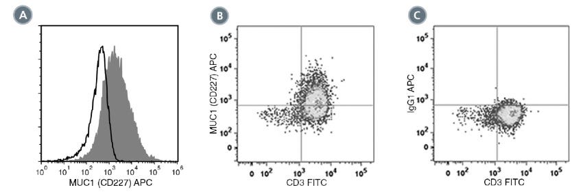
(A) Flow cytometry analysis of human airway epithelial cells cultured in PneumaCult™-ALI Medium at the air-liquid interface. Cells were enzymatically dissociated and labeled with Anti-Human MUC1 (CD227) Antibody, Clone 16A, APC (filled histogram) or Mouse IgG1, kappa Isotype Control Antibody, Clone MOPC-21, APC (Catalog #60070AZ, solid line histogram). (B) Flow cytometry analysis of human peripheral blood lymphocytes following stimulation with phytohemagglutinin (PHA) for 3 days. Cells were labeled with Anti-Human MUC1 (CD227) Antibody, Clone 16A, APC and Anti-Human CD3 Antibody, Clone UCHT1, FITC (Catalog #60011FI). (C) Flow cytometry analysis of human peripheral blood lymphocytes following stimulation with PHA for 3 days. Cells were labeled with Mouse IgG1, kappa Isotype Control Antibody, Clone MOPC-21, APC and Anti-Human CD3 Antibody, Clone UCHT1, FITC.
Find supporting information and directions for use in the Product Information Sheet or explore additional protocols below.
This product is designed for use in the following research area(s) as part of the highlighted workflow stage(s). Explore these workflows to learn more about the other products we offer to support each research area.
Thank you for your interest in IntestiCult™ Organoid Growth Medium (Human). Please provide us with your contact information and your local representative will contact you with a customized quote. Where appropriate, they can also assist you with a(n):
Estimated delivery time for your area
Product sample or exclusive offer
In-lab demonstration
扫描二维码或搜索微信号STEMCELLTech,即可关注我们的微信平台,第一时间接收丰富的技术资源和最新的活动信息。
如您有任何问题,欢迎发消息给STEMCELLTech微信公众平台,或与我们通过电话/邮件联系:400 885 9050 INFO.CN@STEMCELL.COM。


