产品号 #60005_C
Mouse monoclonal IgG1 antibody against human, chimpanzee CD19
The HIB19 antibody reacts with CD19, an ~95 kDa type 1 transmembrane glycoprotein expressed on the surface of B cells throughout all stages of development, from early pre-B cells to plasma cells. Expression is down-regulated but persists in terminally differentiated plasma cells. CD19 is also found on follicular dendritic cells. By associating with CD21 and CD81, CD19 functions as a co-receptor for the B cell receptor and is involved in B cell activation and differentiation. Activation of CD19 is accompanied by phosphorylation of the cytoplasmic domain, which promotes binding to kinases and the induction of intracellular signaling cascades. Mutations in CD19 can result in severe immunodeficiency syndromes.
This antibody clone has been verified for purity assessments of cells isolated with EasySep™ kits, including EasySep™ Human CD19 Positive Selection Kit (Catalog #18054), EasySep™ Human Whole Blood CD19 Positive Selection Kit (Catalog #18084), EasySep™ HLA Whole Blood B Cell Positive Selection Kit (Catalog #18184HLA); partial blocking may be observed, as well as EasySep™ HLA B Cell Enrichment: Complete Processing Kit for Whole Blood (Catalog #19954HLA) and EasySep™ HLA Total Lymphocyte Enrichment: Complete Processing Kit for Whole Blood (Catalog #19961HLA).
Subtype
Primary Antibodies
Target Antigen
CD19
Alternative Names
B4
Reactive Species
Chimpanzee, Human
Conjugation
Alexa Fluor 488, APC, Biotin, FITC, PE, PerCP-Cyanine5.5, Unconjugated
Host Species
Mouse
Cell Type
B Cells
Species
Human, Non-Human Primate
Application
Cell Isolation, CyTOF, Flow Cytometry, Functional Assay, Immunohistochemistry
Area of Interest
Immunology
Clone
HIB19
Gene ID
930
Isotype
IgG1, kappa
Figure 1. Data for Alexa Fluor® 488-Conjugated
(A) Flow cytometry analysis of human peripheral blood mononuclear cells (PBMCs) labeled with Anti-Human CD19 Antibody, Clone HIB19, Alexa Fluor® 488 and anti-human CD45 APC.
(B) Flow cytometry analysis of human PBMCs processed with the EasySep™ Human CD19 Positive Selection Kit and labeled with Anti-Human CD19 Antibody, Clone HIB19, Alexa Fluor® 488. Histograms show labeling of the PBMCs (Start) and isolated cells (Isolated). Labeling with a mouse IgG1, kappa Alexa Fluor® 488 isotype control antibody is shown in the bottom panel (open histogram).
Figure 2. Data for APC-Conjugated
(A) Flow cytometry analysis of human peripheral blood mononuclear cells (PBMCs) labeled with Anti-Human CD19 Antibody, Clone HIB19, APC (filled histogram) or a mouse IgG1, kappa APC isotype control antibody (black line histogram).
(B) Flow cytometry analysis of human PBMCs processed with the EasySep™ Human CD19 Positive Selection Kit and labeled with Anti-Human CD19 Antibody, Clone HIB19, APC. Histograms show labeling of PBMCs (Start) and isolated cells (Isolated). Labeling of start cells with a mouse IgG1, kappa APC isotype control antibody is shown (open histogram).
(C) Flow cytometry analysis of human whole blood nucleated cells processed with the EasySep™ HLA B Cell Enrichment: Complete Processing Kit for Whole Blood and labeled with Anti-Human CD19 Antibody, Clone HIB19, APC. Histograms show labeling of HetaSep™-treated whole blood cells (Start) and isolated cells (Isolated). Labeling of start cells with a mouse IgG1, kappa APC isotype control antibody is shown (open histogram).
Figure 3. Data for Biotin-Conjugated
(A) Flow cytometry analysis of human peripheral blood mononuclear cells (PBMCs) labeled with Anti-Human CD19 Antibody, Clone HIB19, Biotin followed by streptavidin (SAV) APC (filled histogram) or a mouse IgG1, kappa biotin isotype control antibody followed by SAV APC (black line histogram).
(B) Flow cytometry analysis of human PBMCs processed with the EasySep™ Human CD19 Positive Selection Kit and labeled with Anti-Human CD19 Antibody, Clone HIB19, Biotin followed by streptavidin (SAV) APC. Histograms show labeling of PBMCs (Start) and isolated cells (Isolated). Labeling of start cells with a mouse IgG1, kappa biotin isotype control antibody followed by SAV APC is shown (open histogram).
(C) Flow cytometry analysis of human buffy coat nucleated cells processed with the EasySep™ HLA Whole Blood B Cell Positive Selection Kit and labeled with Anti-Human CD19 Antibody, Clone HIB19, Biotin followed by streptavidin (SAV) APC. Histograms show labeling of buffy coat nucleated cells (Start) and isolated cells (Isolated). Labeling of start cells with a mouse IgG1, kappa biotin isotype control antibody followed by SAV APC is shown (open histogram).
Figure 4. Data for FITC-Conjugated
(A) Flow cytometry analysis of human peripheral blood mononuclear cells (PBMCs) labeled with Anti-Human CD19 Antibody, Clone HIB19, FITC (filled histogram) or a mouse IgG1, kappa FITC isotype control antibody (black line histogram).
(B) Flow cytometry analysis of human PBMCs processed with the EasySep™ Human CD19 Positive Selection Kit and labeled with Anti-Human CD19 Antibody, Clone HIB19, FITC. Histograms show labeling of PBMCs (Start) and isolated cells (Isolated). Labeling of start cells with a mouse IgG1, kappa FITC isotype control antibody is shown (open histogram).
(C) Flow cytometry analysis of human buffy coat nucleated cells processed with the EasySep™ Human Whole Blood CD19 Positive Selection Kit and labeled with Anti-Human CD19 Antibody, Clone HIB19, FITC. Histograms show labeling of buffy coat nucleated cells (Start) and isolated cells (Isolated). Labeling of start cells with a mouse IgG1, kappa FITC isotype control antibody is shown (open histogram).
Figure 5. Data for PE-Conjugated
(A) Flow cytometry analysis of human peripheral blood mononuclear cells (PBMCs) labeled with Anti-Human CD19 Antibody, Clone HIB19, PE and antihuman CD45 APC.
(B) Flow cytometry analysis of human PBMCs processed with the EasySep™ Human CD19 Positive Selection Kit and labeled with Anti-Human CD19 Antibody, Clone HIB19, PE. Histograms show labeling of the PBMCs (Start) and isolated cells (Isolated). Labeling with a mouse IgG1, kappa PE isotype control antibody is shown in the bottom panel (open histogram).
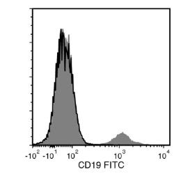
Figure 6. Data for Unconjugated
Flow cytometry analysis of human peripheral blood mononuclear cells (PBMCs) labeled with Anti-Human CD19 Antibody, Clone HIB19, followed by Goat Anti-Mouse IgG (H+L) Antibody, Polyclonal, FITC (Catalog #60138FI; filled histogram) or a mouse IgG1, kappa isotype control antibody followed by Goat Anti-Mouse IgG (H+L) Antibody, Polyclonal, FITC (solid line histogram).
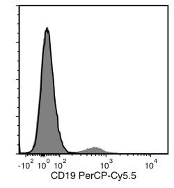
Figure 7. Data for PerCP-Cy55-Conjugated
Flow cytometry analysis of human peripheral blood mononuclear cells (PBMCs) labeled with Anti-Human CD19 Antibody, Clone HIB19, PerCP-Cy5.5 (filled histogram) or a mouse IgG1, kappa PerCP-Cy5.5 isotype control antibody (solid line histogram).
Find supporting information and directions for use in the Product Information Sheet or explore additional protocols below.
This product is designed for use in the following research area(s) as part of the highlighted workflow stage(s). Explore these workflows to learn more about the other products we offer to support each research area.
Thank you for your interest in IntestiCult™ Organoid Growth Medium (Human). Please provide us with your contact information and your local representative will contact you with a customized quote. Where appropriate, they can also assist you with a(n):
Estimated delivery time for your area
Product sample or exclusive offer
In-lab demonstration
扫描二维码或搜索微信号STEMCELLTech,即可关注我们的微信平台,第一时间接收丰富的技术资源和最新的活动信息。
如您有任何问题,欢迎发消息给STEMCELLTech微信公众平台,或与我们通过电话/邮件联系:400 885 9050 INFO.CN@STEMCELL.COM。