产品号 #60039_C
Mouse monoclonal IgG1 antibody against human, mouse CD105 (endoglin)
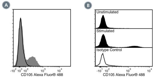
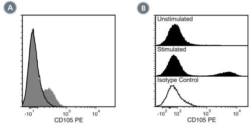
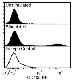
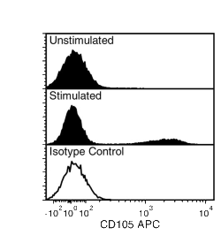
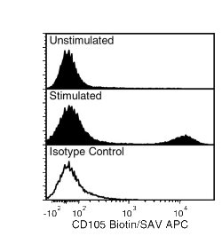
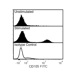
The 43A3 antibody reacts with CD105 (endoglin), an ~180 kDa cell surface glycoprotein which is a disulfide-bonded homodimer of ~90 kDa type I transmembrane subunits. CD105 is a component of the TGF-β receptor complex and is expressed by vascular endothelial smooth muscle cells, syncytiotrophoblasts of placenta and activated macrophages, and at relatively low levels by stromal fibroblasts. Its expression is also observed in some types of tumors, and levels are up-regulated on the endothelium during angiogenesis. In concert with signaling receptors, CD105 binds to TGF-β1 and TGF-β3 with high affinity, but does not bind TGF-β2. Other ligands reportedly include Activin A, BMP-2, and BMP-7. CD105 has important roles in angiogenesis, cardiovascular development, and vascular remodeling, and the protein serves a regulatory role in cytoskeletal reorganization by modulating the sites of focal adhesion and cellular migration. Certain mutations in CD105 result in the autosomal dominant disorder hereditary hemorrhagic telangiectasia.
This antibody clone has been verified for labeling human mesenchymal cells grown in MesenCult™ Proliferation Kit (Human; Catalog #05411) and MesenCult™-XF Medium (Catalog #05420).
Subtype
Primary Antibodies
Target Antigen
CD105 (Endoglin)
Alternative Names
Endoglin
Reactive Species
Human, Mouse
Conjugation
Alexa Fluor 488, APC, Biotin, FITC, PE, Unconjugated
Host Species
Mouse
Cell Type
Hematopoietic Stem and Progenitor Cells, Mesenchymal Stem and Progenitor Cells
Species
Human, Mouse
Application
Flow Cytometry, Immunoprecipitation, Western Blotting
Area of Interest
Endothelial Cell Biology, Stem Cell Biology
Clone
43A3
Gene ID
2022
Isotype
IgG1, kappa
Figure 1. Data for Alexa Fluor® 488-Conjugated
(A) Flow cytometry analysis of human peripheral blood mononuclear cells (PBMCs) labeled with Anti-Human CD105 Antibody, Clone 43A3, Alexa Fluor® 488 (filled histogram) or a mouse IgG1, kappa Alexa Fluor® 488 isotype control antibody (solid line histogram). (B) Flow cytometry analysis of human PBMCs cultured for 24 hours with or without lipopolysaccharide (LPS) and labeled with Anti-Human CD105 Antibody, Clone 43A3, Alexa Fluor® 488. Histograms show labeling of PBMCs cultured in the absence (Unstimulated) or presence (Stimulated) of LPS. Labeling of LPS-stimulated PBMCs with a mouse IgG1, kappa isotype control antibody, Alexa Fluor® 488 is shown (solid line histogram).
Figure 2. Data for PE-Conjugated
(A) Flow cytometry analysis of human peripheral blood mononuclear cells (PBMCs) labeled with Anti-Human CD105 Antibody, Clone 43A3, PE (filled histogram) or a mouse IgG1, kappa PE isotype control antibody (solid line histogram). (B) Flow cytometry analysis of human PBMCs cultured for 24 hours with or without lipopolysaccharide (LPS) and labeled with Anti-Human CD105 Antibody, Clone 43A3, PE. Histograms show labeling of PBMCs cultured in the absence (Unstimulated) or presence (Stimulated) of LPS. Labeling of LPS-stimulated PBMCs with a mouse IgG1, kappa isotype control antibody, PE is shown (solid line histogram).
Figure 3. Data for Unconjugated
Flow cytometry analysis of human peripheral blood mononuclear cells (PBMCs) cultured for 24 hours with or without lipopolysaccharide (LPS) and labeled with Anti-Human CD105 Antibody, Clone 43A3, followed by a rat anti-mouse IgG1 antibody, PE. Histograms show labeling of PBMCs cultured in the absence (Unstimulated) or presence (Stimulated) of LPS. Labeling of LPS-stimulated PBMCs with a mouse IgG1, kappa isotype control antibody, followed by a rat anti-mouse IgG1 antibody, PE is shown (solid line histogram).
Figure 4. Data for APC-Conjugated
Flow cytometry analysis of human peripheral blood mononuclear cells (PBMCs) cultured for 24 h with or without lipopolysaccharide (LPS) and labeled with Anti-Human CD105 Antibody, Clone 43A3, APC. Histograms show labeling of PBMCs cultured in the absence (Unstimulated) or presence (Stimulated) of LPS. Labeling of LPS-stimulated PBMCs with a mouse IgG1, kappa isotype control antibody, APC is shown in the bottom panel (solid line histogram).
Figure 5. Data for Biotin-Conjugated
Flow cytometry analysis of human peripheral blood mononuclear cells (PBMCs) cultured for 24 h with or without lipopolysaccharide (LPS) and labeled with Anti-Human CD105 Antibody, Clone 43A3, Biotin followed by streptavidin (SAV) APC. Histograms show labeling of PBMCs cultured in the absence (Unstimulated) or presence (Stimulated) of LPS. Labeling of LPS-stimulated PBMCs with a biotinylated mouse IgG1, kappa isotype control antibody followed by SAV APC is shown in the bottom panel (solid line histogram).
Figure 6. Data for FITC-Conjugated
Flow cytometry analysis of human peripheral blood mononuclear cells (PBMCs) cultured for 24 h with or without lipopolysaccharide (LPS) and labeled with Anti-Human CD105 Antibody, Clone 43A3, FITC. Histograms show labeling of PBMCs cultured in the absence (Unstimulated) or presence (Stimulated) of LPS. Labeling of LPS-stimulated PBMCs with a mouse IgG1, kappa isotype control antibody, FITC is shown in the bottom panel (solid line histogram).
Find supporting information and directions for use in the Product Information Sheet or explore additional protocols below.
This product is designed for use in the following research area(s) as part of the highlighted workflow stage(s). Explore these workflows to learn more about the other products we offer to support each research area.
Thank you for your interest in IntestiCult™ Organoid Growth Medium (Human). Please provide us with your contact information and your local representative will contact you with a customized quote. Where appropriate, they can also assist you with a(n):
Estimated delivery time for your area
Product sample or exclusive offer
In-lab demonstration
扫描二维码或搜索微信号STEMCELLTech,即可关注我们的微信平台,第一时间接收丰富的技术资源和最新的活动信息。
如您有任何问题,欢迎发消息给STEMCELLTech微信公众平台,或与我们通过电话/邮件联系:400 885 9050 INFO.CN@STEMCELL.COM。