产品号 #100-0328_C
A cellular protein for detection of apoptotic cells
Annexin V is a member of the annexin family of proteins that bind to membrane phospholipids in the presence of calcium. This dye has high affinity for phosphatidylserine (PS) that is present in the inner leaflet of the plasma membrane. During early-stage cell apoptosis, PS is translocated from the inner to the outer leaflet of the plasma membrane, exposing it to the external environment. Annexin V, a characteristic marker for early cell apoptosis, detects the translocation of PS to the external environment.
Annexin V is used along with viability dyes such as 7-AAD (Catalog #75001) or Propidium Iodide (Catalog #75002). The process of PS translocation occurs prior to the loss of membrane integrity. Therefore, as cells progress through apoptosis and towards necrosis, the cell membrane is compromised and consequently, viability dyes pass into the cell. Thus, cells undergoing early apoptosis stain positive for Annexin V and negative for viability dyes, while apoptotic death or necrosis is characterized by positive staining for both Annexin V and the viability dye.
Conjugation
APC, FITC, PE
Application
Flow Cytometry
Area of Interest
Immunology
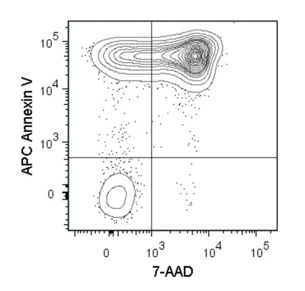
Figure 1. Data for Annexin V, APC
Flow cytometry analysis of C57BL/6 mouse thymocytes incubated at 37°C with 1 µM dexamethasone overnight. Cells were harvested and labeled with APC-conjugated Annexin V and 7-AAD (Catalog #75001).
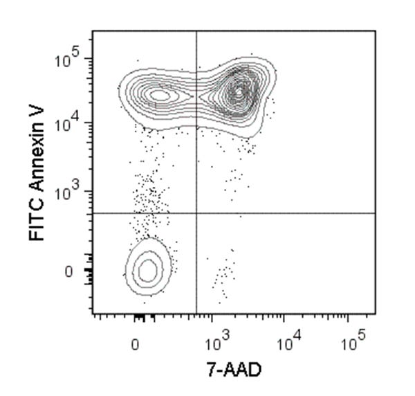
Figure 2. Data for Annexin V, FITC
Figure showing flow cytometry analysis of C57BL/6 mouse thymocytes incubated at 37°C with 1 µM dexamethasone overnight. Cells were harvested and labeled with FITC Annexin V and 7-AAD.
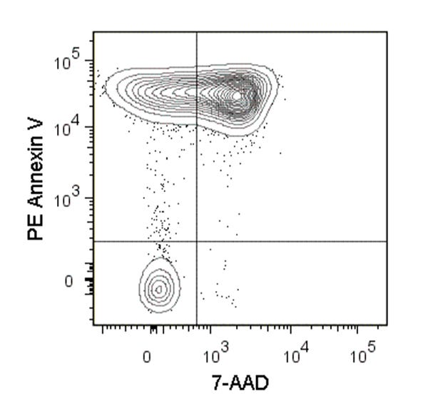
Figure 3. Data for Annexin V, PE
Figure showing flow cytometry analysis of C57BL/6 mouse thymocytes incubated at 37°C with 1 µM dexamethasone overnight. Cells were harvested and labeled with PE Annexin V and 7-AAD.
Find supporting information and directions for use in the Product Information Sheet or explore additional protocols below.
This product is designed for use in the following research area(s) as part of the highlighted workflow stage(s). Explore these workflows to learn more about the other products we offer to support each research area.
Thank you for your interest in IntestiCult™ Organoid Growth Medium (Human). Please provide us with your contact information and your local representative will contact you with a customized quote. Where appropriate, they can also assist you with a(n):
Estimated delivery time for your area
Product sample or exclusive offer
In-lab demonstration
扫描二维码或搜索微信号STEMCELLTech,即可关注我们的微信平台,第一时间接收丰富的技术资源和最新的活动信息。
如您有任何问题,欢迎发消息给STEMCELLTech微信公众平台,或与我们通过电话/邮件联系:400 885 9050 INFO.CN@STEMCELL.COM。