StemSpan™巨核细胞扩增补充(100X)

人巨核细胞扩增的无血清培养补充物

产品号 #(选择产品)

产品号 #02696_C

人巨核细胞扩增的无血清培养补充物

产品优势

  • 配制成在CD34+ CB或BM细胞启动的液体培养中产生大量的人巨核细胞。
  • 针对StemSpan™媒体进行了优化。当与StemSpan™SFEM II结合使用时,与市场上其他无血清培养基相比,可支持高达2倍的人CD34+ CB细胞的巨核细胞扩增。
  • 作为100X浓缩物供应。在解冻和混合后,试管内容物可以直接添加到任何选择的造血细胞扩增培养基中。

概述

StemSpan™巨核细胞扩增补充剂(100X)包含重组人细胞因子的组合,用于选择性地促进从人脐带血(CB)或骨髓(BM)样本中分离的CD34+细胞的人巨核细胞祖细胞的扩增和分化。

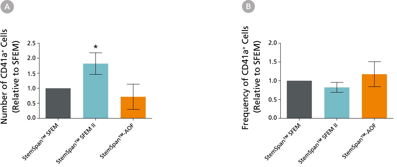
StemSpan™巨核细胞扩增补充(100X)用于与以下任何StemSpan™培养基结合使用:
•StemSpan™SFEM(目录#09600)
StemSpan™SFEM II(目录#09605)
StemSpan™-XF(目录#100-0073)
StemSpan™-AOF(目录#100-0130)

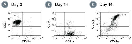
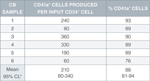
当添加到无血清培养基中时,StemSpan™巨核细胞扩增补充剂通常促进每个输入CD34+细胞在CD34+人CB细胞启动的14天液体培养中产生数百个巨核细胞。请参阅data选项卡了解更多详细信息。

Contains
• Recombinant human stem cell factor (SCF)
• Recombinant human interleukin 6 (IL-6)
• Recombinant human interleukin 9 (IL-9)
• Recombinant human thrombopoietin (TPO)
 
Subtype
Supplements
 
Cell Type
Hematopoietic Stem and Progenitor Cells, Megakaryocytes
 
Species
Human
 
Application
Cell Culture, Differentiation, Expansion
 
Brand
StemSpan
 
Area of Interest
Stem Cell Biology, Transplantation Research
 
Formulation Category
Serum-Free
 

Data Figures

Production of megakaryocytes by expansion and lineage-specific differentiation of CD34+ human cord blood cells cultured in StemSpan™ SFEM containing Megakaryocyte Expansion Supplement

Figure 1. Production of Megakaryocytes by Expansion and Lineage-Specific Differentiation of CD34+ Human Cord Blood Cells Cultured in StemSpan™ SFEM Containing Megakaryocyte Expansion Supplement

Flow cytometry dot plots showing expression of the hematopoietic stem and progenitor cell marker CD34 and megakaryocyte markers CD41a and CD42b (A) before and (B,C) after culture of CD34+ cord blood cells for 14 days in StemSpan™ SFEM containing Megakaryocyte Expansion Supplement. The frequency of CD34+ cells declined from 90% before culture to <3% after 14 days, in parallel with a gradual accumulation of CD41a+CD42b+ megakaryocytes from 80% before and after culture, respectively.

Table 1. Production of Megakaryocytes from CD34+ Human Cord Blood Cells Cultured in StemSpan™ SFEM Containing Megakaryocyte Expansion Supplement

Production of megakaryocytes from CD34+ human cord blood cells cultured in StemSpan™ SFEM containing Megakaryocyte Expansion Supplement

Numbers and percent of CD41a+ cells produced after 14 days of culture of enriched CD34+ cells from 6 independent cord blood (CB) samples. *95% confidence limits, the range within which 95% of the results will typically fall.

Comparison of megakaryocyte expansion in different StemSpan™ media containing Megakaryocyte Expansion Supplement

Figure 2. Comparison of Megakaryocyte Expansion in Different StemSpan™ Media Containing Megakaryocyte Expansion Supplement

(A) Average numbers and (B) frequencies of CD41+ megakaryocytic cells normalized relative to the values obtained in StemSpan™ SFEM (grey bars) after culturing purified CD34+ cord blood cells (n=6) for 14 days in StemSpan™ SFEM, SFEM II (blue bars) and AOF (orange bars) media containing Megakaryocyte Expansion Supplement. Vertical lines indicate 95% confidence limits, the range within which 95% of results typically fall. *The numbers of CD41a+ cells were significantly higher in SFEM II (p<0.01, paired t-test, n=6) compared to SFEM and AOF medium.

Note: Data for StemSpan™-AOF shown were generated with the original phenol red-containing version StemSpan™-ACF (Catalog #09855). However internal testing showed that the performance of the new phenol red-free, cGMP-manufactured version, StemSpan™-AOF (Catalog #100-0130) was comparable.

Protocols and Documentation

Find supporting information and directions for use in the Product Information Sheet or explore additional protocols below.

Document Type
Product Name
Catalog #
Lot #
Language
Catalog #
02696
Lot #
All
Language
English
Document Type
Safety Data Sheet
Catalog #
02696
Lot #
All
Language
English

Applications

This product is designed for use in the following research area(s) as part of the highlighted workflow stage(s). Explore these workflows to learn more about the other products we offer to support each research area.

Resources and Publications

Educational Materials (7)

Publications (11)

Identification of unipotent megakaryocyte progenitors in human hematopoiesis. Miyawaki K et al. Blood 2017 MAR

Abstract

The developmental pathway for human megakaryocytes remains unclear and the definition of pure unipotent megakaryocyte progenitor is still controversial. Using single-cell transcriptome analysis, we have identified a cluster of cells within immature hematopoietic stem and progenitor cell populations that specifically express genes related to the megakaryocyte lineage. We used CD41 as a positive marker to identify these cells within the CD34(+)CD38(+)IL-3Rα(dim)CD45RA(-) common myeloid progenitor (CMP) population. These cells lacked erythroid and granulocyte/macrophage potential, but exhibited robust differentiation into the megakaryocyte lineage at a high frequency, both in vivo and in vitro The efficiency and expansion potential of these cells exceeded those of conventional bipotent megakaryocyte/erythrocyte progenitors. Accordingly, the CD41(+) CMP was defined as a unipotent megakaryocyte progenitor (MegP) that is likely to represent the major pathway for human megakaryopoiesis, independent of canonical megakaryocyte-erythroid lineage bifurcation. In the bone marrow of patients with essential thrombocythemia, the MegP population was significantly expanded in the context of a high burden of Janus kinase 2 mutations. Thus, the prospectively isolatable and functionally homogeneous human MegP will be useful for the elucidation of the mechanisms underlying normal and malignant human hematopoiesis.
miR-34a contributes to megakaryocytic differentiation of K562 cells independently of p53. Navarro F et al. Blood 2009 SEP

Abstract

The role of miRNAs in regulating megakaryocyte differentiation was examined using bipotent K562 human leukemia cells. miR-34a is strongly up-regulated during phorbol ester-induced megakaryocyte differentiation, but not during hemin-induced erythrocyte differentiation. Enforced expression of miR-34a in K562 cells inhibits cell proliferation, induces cell-cycle arrest in G(1) phase, and promotes megakaryocyte differentiation as measured by CD41 induction. miR-34a expression is also up-regulated during thrombopoietin-induced differentiation of CD34(+) hematopoietic precursors, and its enforced expression in these cells significantly increases the number of megakaryocyte colonies. miR-34a directly regulates expression of MYB, facilitating megakaryocyte differentiation, and of CDK4 and CDK6, to inhibit the G(1)/S transition. However, these miR-34a target genes are down-regulated rapidly after inducing megakaryocyte differentiation before miR-34a is induced. This suggests that miR-34a is not responsible for the initial down-regulation but may contribute to maintaining their suppression later on. Previous studies have implicated miR-34a as a tumor suppressor gene whose transcription is activated by p53. However, in p53-null K562 cells, phorbol esters induce miR-34a expression independently of p53 by activating an alternative phorbol ester-responsive promoter to produce a longer pri-miR-34a transcript.
In vitro expanded cells contributing to rapid severe combined immunodeficient repopulation activity are CD34+38-33+90+45RA-. Vanheusden K et al. Stem cells (Dayton, Ohio) 2007 JAN

Abstract

Expansion of hematopoietic stem cells could be used clinically to shorten the prolonged aplastic phase after umbilical cord blood (UCB) transplantation. In this report, we investigated rapid severe combined immunodeficient (SCID) repopulating activity (rSRA) 2 weeks after transplantation of CD34(+) UCB cells cultured with serum on MS5 stromal cells and in serum- and stroma-free cultures. Various subpopulations obtained after culture were studied for rSRA. CD34(+) expansion cultures resulted in vast expansion of CD45(+) and CD34(+) cells. Independent of the culture method, only the CD34(+)33(+)38(-) fraction of the cultured cells contained rSRA. Subsequently, we subfractionated the CD34(+)38(-) fraction using stem cell markers CD45RA and CD90. In vitro differentiation cultures showed CD34(+) expansion in both CD45RA(-) and CD90(+) cultures, whereas little increase in CD34(+) cells was observed in both CD45RA(+) and CD90(-) cultures. By four-color flow cytometry, we could demonstrate that CD34(+)38(-)45RA(-) and CD34(+)38(-)90(+) cell populations were largely overlapping. Both populations were able to reconstitute SCID/nonobese diabetic mice at 2 weeks, indicating that these cells contained rSRA activity. In contrast, CD34(+)38(-)45RA(+) or CD34(+)38(-)90(-) cells contributed only marginally to rSRA. Similar results were obtained when cells were injected intrafemorally, suggesting that the lack of reconstitution was not due to homing defects. In conclusion, we show that after in vitro expansion, rSRA is mediated by CD34(+)38(-)90(+)45RA(-) cells. All other cell fractions have limited reconstitutive potential, mainly because the cells have lost stem cell activity rather than because of homing defects. These findings can be used clinically to assess the rSRA of cultured stem cells.

更多信息

更多信息
Species Human
Contains • Recombinant human stem cell factor (SCF) • Recombinant human interleukin 6 (IL-6) • Recombinant human interleukin 9 (IL-9) • Recombinant human thrombopoietin (TPO)
Formulation Category Serum-Free
PRODUCTS ARE FOR RESEARCH USE ONLY AND NOT INTENDED FOR HUMAN OR ANIMAL DIAGNOSTIC OR THERAPEUTIC USES UNLESS OTHERWISE STATED. FOR ADDITIONAL INFORMATION ON QUALITY AT STEMCELL, REFER TO WWW.STEMCELL.COM/COMPLIANCE.
版权 © 2025 STEMCELL Technologies 技术有限公司。保留所有权利。