STEMdiff™ 肾类器官试剂盒

用于从人多能干细胞(hPSCs)培养肾类器官的无血清培养基试剂盒

产品号 #(选择产品)

产品号 #05160_C

用于从人多能干细胞(hPSCs)培养肾类器官的无血清培养基试剂盒

产品优势

  • 相关性强:可生成模拟发育中肾单位及相关内皮和间充质的人类肾类器官。
  • 操作简便:采用两阶段培养体系和简明易懂的操作流程,最大限度减少培养操作。
  • 可靠性高:通过优化配方和严格的质量控制,降低实验差异性。
  • 高通量:可在96孔和384孔板中重复生成类器官。

产品组分包括

  • STEMdiff™ 肾脏基础培养基,100 mL
  • STEMdiff™ 肾脏补充剂 SG (100X),200 µL
  • STEMdiff™ 肾脏补充剂 DM (50X),1.6 mL
Need a high-quality cell source? Use the hiPSC SCTi004-A (male) control line, manufactured with mTeSR™ Plus.

What Our Scientist Says

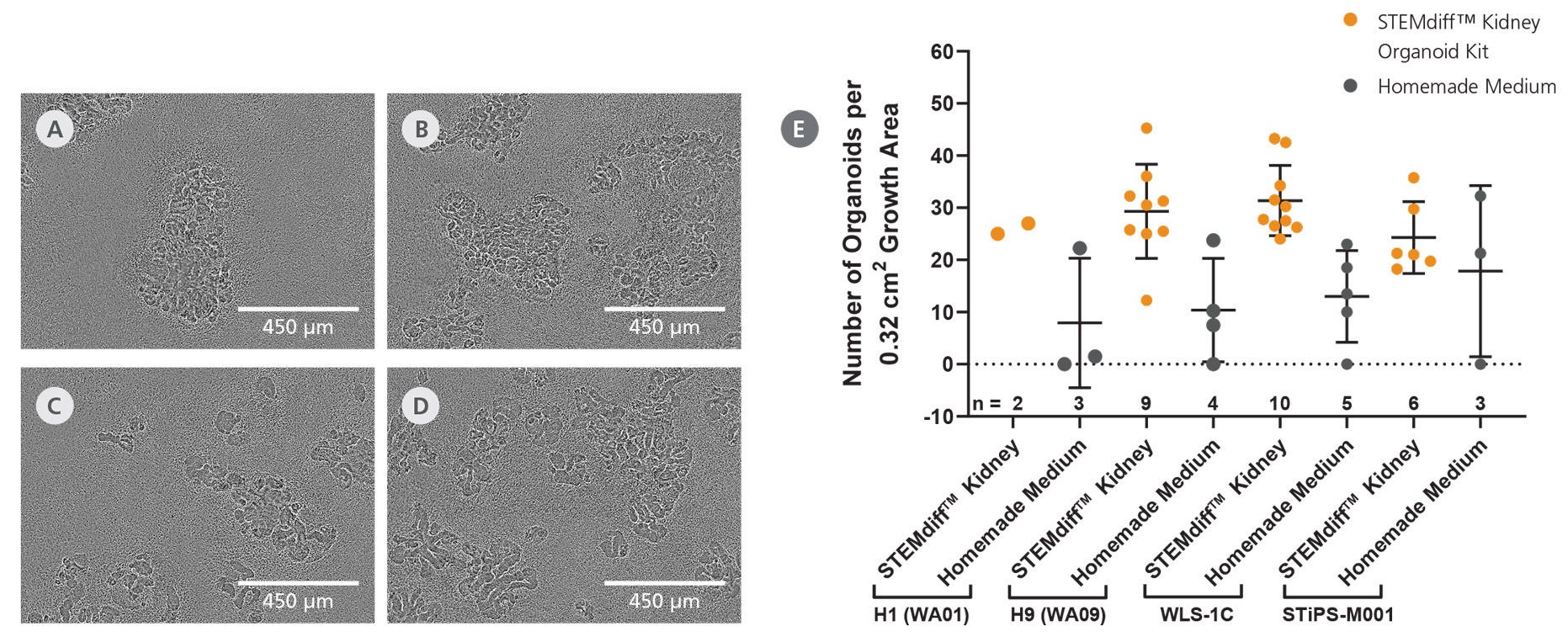
We developed the STEMdiff™ Kidney Organoid Kit to enable researchers to easily generate kidney organoids with a typical nephron-like segmentation that are suitable for disease modeling, nephrotoxicity assessment, and other applications.

Philipp KramerSenior Scientist
Philipp Kramer, Senior Scientist

总览

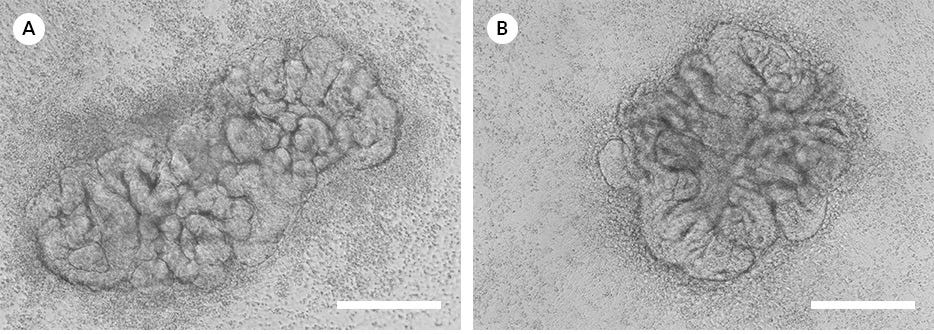
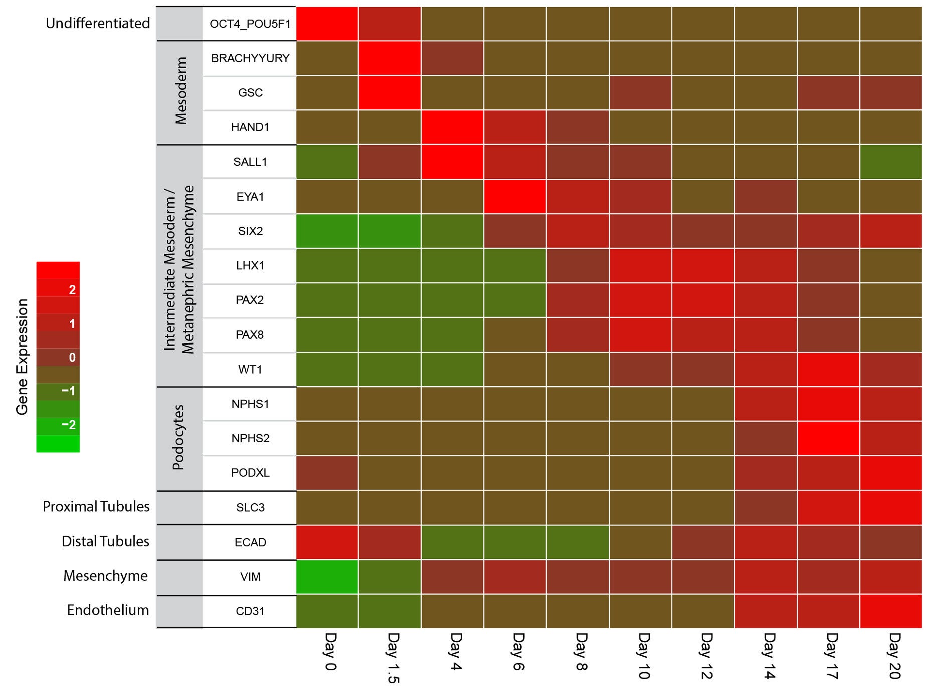
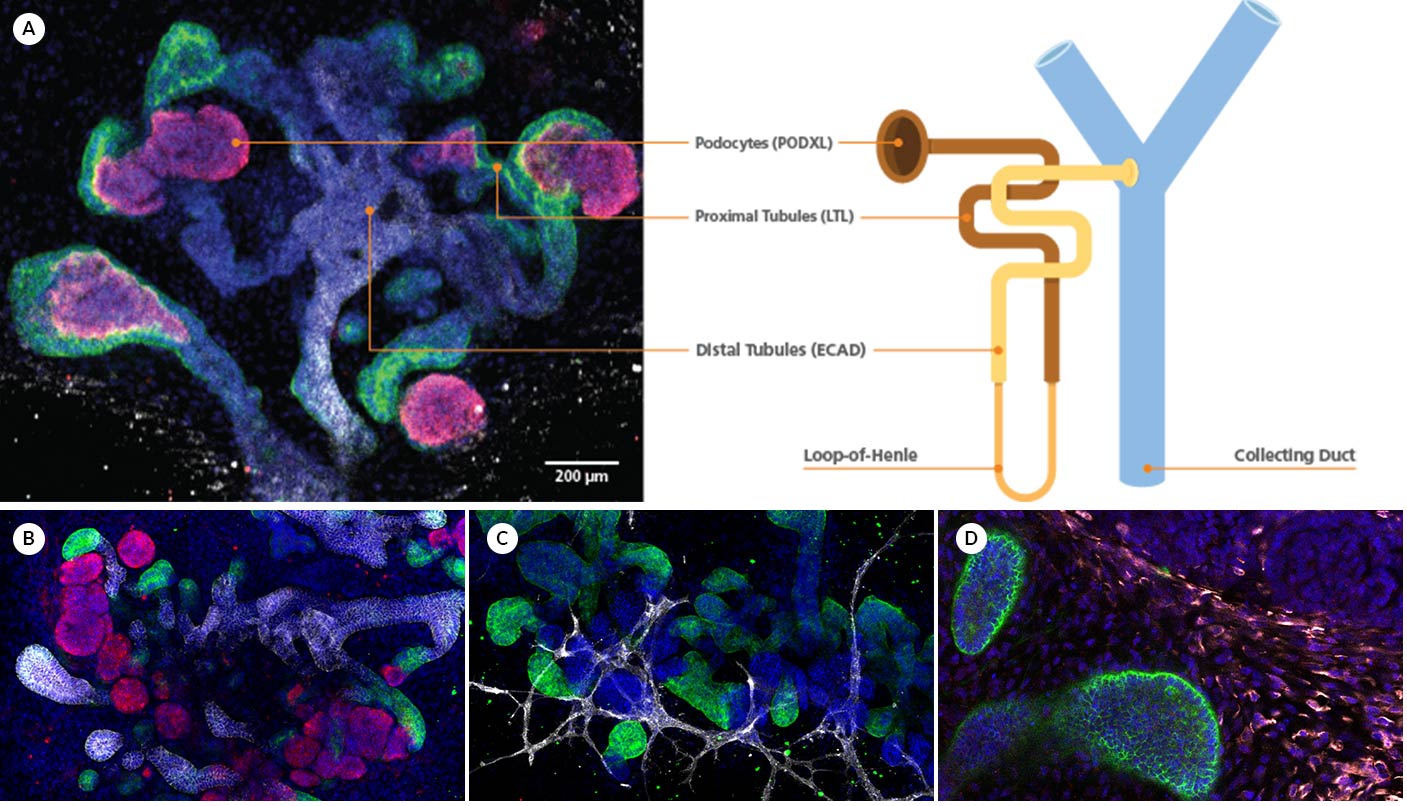
STEMdiff™ 肾脏类器官试剂盒是一套完整的无血清细胞培养系统,可在简便的两阶段分化流程中高效且可重复地生成源自人类多能干细胞(hPSC)的肾脏类器。这些肾脏类器官由足细胞、近端小管、远端小管以及相关的内皮细胞和间充质组成。

使用 STEMdiff™ 肾脏类器官试剂盒生成的肾脏类器官已通过表型高通量分析(例如肾毒性化合物筛选)的兼容性测试。该类器官模型同样适用于发育生物学研究和疾病模型构建,提供了一个相关性强且便捷的研究平台。

该试剂盒已针对先前在 mTeSR1(目录号 #85850)中培养的 hPSC 的分化进行了优化。对于使用 mTeSR Plus(目录号 #100-0276)维持的 hPSC,也可兼容使用,但在实验流程的前3天必须使用 mTeSR1 培养基。

包含
Serum-free medium kit for the culture of kidney orgaonids from hPSCs
 
亚型
专用培养基
 
细胞类型
肾细胞,中胚层,PSC衍生
 
种属

 
应用
细胞培养,分化,功能学筛选,类器官培养
 
品牌
STEMdiff
 
研究领域
疾病建模,药物发现和毒理检测,上皮细胞研究,类器官
 
制剂类别
无血清
 

Data Figures

Figure 1. Representative Images of Kidney Organoids

The STEMdiff™ Kidney Organoid Kit facilitates the directed differentiation of hPSCs to form kidney organoids that model the developing nephron. Pictured are human kidney organoids grown using the STEMdiff™ Kidney Organoid Kit differentiated from (A) iPS (WLS-1C) or (B) ES (H9) cells and imaged on day 12 and day 18 of differentiation, respectively.

Figure 2. Schematic for Differentiation from hPSCs to Human Kidney Organoids with the STEMdiff™ Kidney Organoid Kit

hPSC cultures progress through a simple two-stage process to generate kidney organoids. hPSCs are first plated and overlaid with Corning® Matrigel® to form cavitated spheroids. On the following day (Day 0), differentiation of cavitated PSC spheroids is initiated by changing medium from mTeSR™1 to STEMdiff™ Kidney Organoid Kit. During the next 18 days, the cells are directed through stages of the late primitive streak, intermediate mesoderm, and metanephric mesoderm to give rise to kidney organoids that are composed of podocytes, proximal and distal tubules, and an associated endothelium and mesenchyme.

Figure 3. Efficient Differentiation of Human Pluripotent Stem Cells into Self-Organizing Kidney Organoids

The STEMdiff™ Kidney Organoid Kit enables high-efficiency generation of kidney organoids from both ES (A, H1; B, H9) and iPS (C, WLS-1C; D, STiPS-M001) cell lines. (E) Kidney organoids were grown using the STEMdiff™ Kidney Organoid Kit or using home-made medium, and the average number of organoids per well of a 96-well plate was quantified on Day 18. All four tested cell lines were capable of differentiating into self-organizing kidney organoids that form convoluted tubular structures with high efficiency (mean ± SD, n ≥ 2).

Figure 4. Kidney Organoids Grown Using the STEMdiff™ Kidney Organoid Kit Show the Expected Changes in Gene Expression During Differentiation

As organoids progress through the stages of differentiation to more mature renal cell types, gene expression shifts from markers of pluripotency (day 0) to the mesoderm (day 1.5 - 4) and to the intermediate mesoderm/metanephric mesenchyme by day 4 - 12. Markers of the podocytes, proximal and distal tubules, mesenchyme and endothelium are observable starting by day 14. Marker levels were assessed in four independent experiments by RT-qPCR and normalized to expression levels of undifferentiated cells.

Figure 5. Kidney Organoids form Convoluted Tubular Structures with Typical Nephron-like Segmentation

(A) During differentiation, kidney organoids form convoluted tubular structures that resemble the structure and segmentation of the developing nephron. (B) These organoids express markers of the renal epithelium including podocyte marker podocalyxin (red), proximal tubule marker lotus tetragonolobus lectin (green), and distal tubule marker E-cadherin (white). Cultures also contain (C) endothelial cells stained by platelet endothelial cell adhesion molecule (CD31, white), as well as (D) mesenchymal markers vimentin (white) and Meis homeobox family (MEIS1/2/3, red). Images (C) and (D) are co-stained for proximal tubule marker lotus tetragonolobus lectin (green). All images show nuclei stained with DAPI (blue).

Protocols and Documentation

Find supporting information and directions for use in the Product Information Sheet or explore additional protocols below.

Document Type
Product Name
Catalog #
Lot #
Language
Catalog #
05160
Lot #
All
Language
English
Document Type
Technical Manual
Catalog #
05160
Lot #
All
Language
English
Document Type
Safety Data Sheet 1
Catalog #
05160
Lot #
All
Language
English
Document Type
Safety Data Sheet 2
Catalog #
05160
Lot #
All
Language
English
Document Type
Safety Data Sheet 3
Catalog #
05160
Lot #
All
Language
English

Applications

This product is designed for use in the following research area(s) as part of the highlighted workflow stage(s). Explore these workflows to learn more about the other products we offer to support each research area.

Resources and Publications

Educational Materials (19)

更多信息

更多信息
Species Human
Contains Serum-free medium kit for the culture of kidney orgaonids from hPSCs
Formulation Category Serum-Free
PRODUCTS ARE FOR RESEARCH USE ONLY AND NOT INTENDED FOR HUMAN OR ANIMAL DIAGNOSTIC OR THERAPEUTIC USES UNLESS OTHERWISE STATED. FOR ADDITIONAL INFORMATION ON QUALITY AT STEMCELL, REFER TO WWW.STEMCELL.COM/COMPLIANCE.
版权 © 2025 STEMCELL Technologies 技术有限公司。保留所有权利。