产品号 #05010_C
人PSCs向心室心肌细胞分化的无血清培养基和人PSCs来源的心肌细胞的长期维持
人PSCs向心室心肌细胞分化的无血清培养基和人PSCs来源的心肌细胞的长期维持
cGMP, enzyme-free cell dissociation reagent
Dulbecco’s phosphate-buffered saline without calcium and magnesium
RHO/ROCK pathway inhibitor; Inhibits ROCK1 and ROCK2
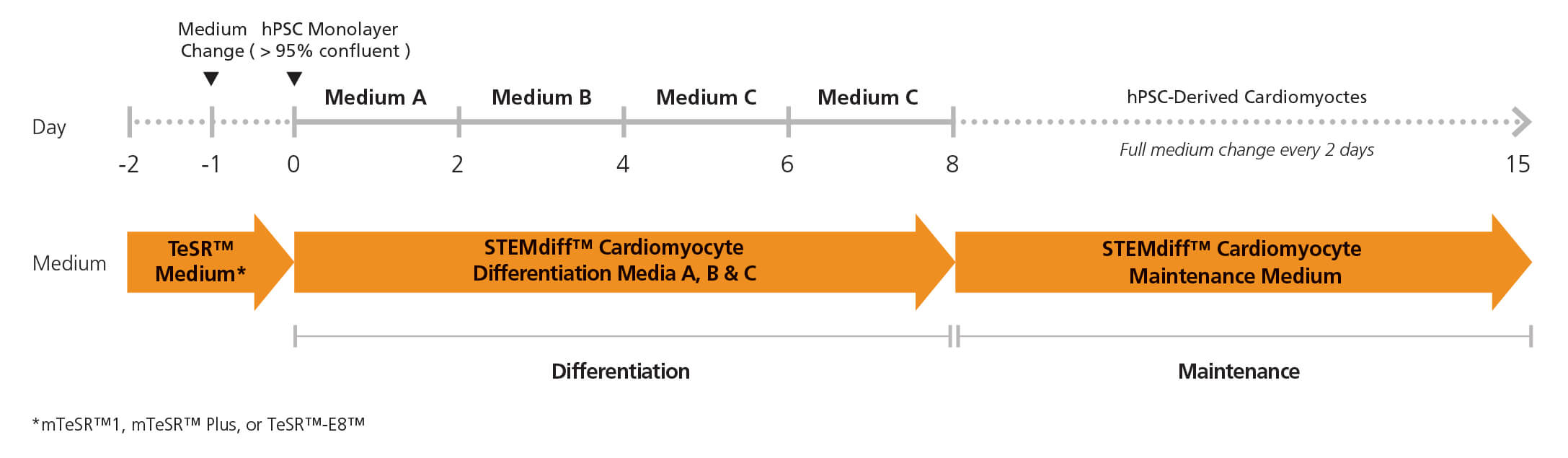
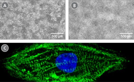
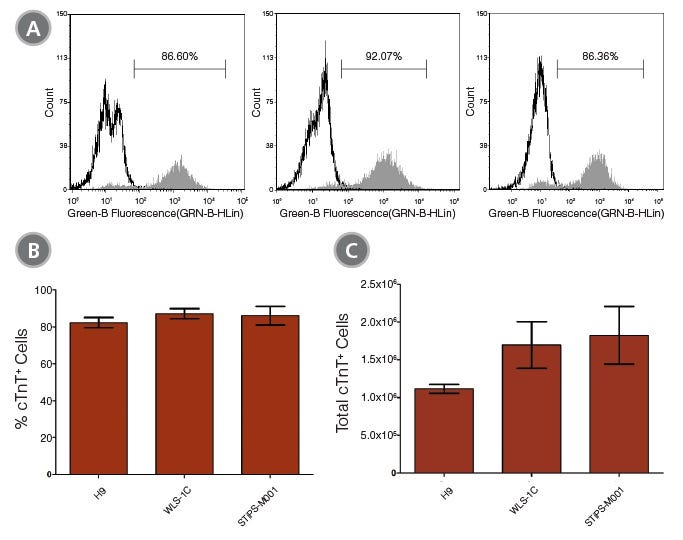
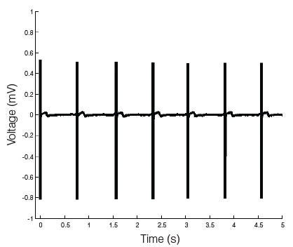
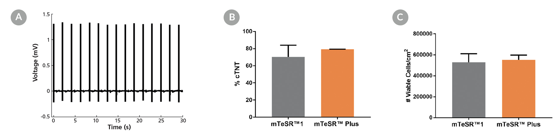
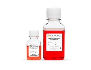
STEMdiff™心室心肌细胞分化试剂盒(目录#05010)包括用于将人胚胎干(ES)和诱导多能干细胞(iPS)细胞(人多能干细胞[hPSCs])分化为心室心肌细胞(心肌肌钙蛋白t阳性[cTnT+])的培养基,以及用于维持hpsc来源的心肌细胞的培养基。该无血清试剂盒可用于生成源自mTeSR™1(目录#85850),mTeSR™Plus(目录#100-0276),TeSR™-AOF(目录#100-0401)或TeSR™-E8™(目录#05990)中维持的hPSCs的团块培养的心室心肌细胞。这些细胞中超过80%为cTnT+。12孔板的单孔平均可收获1 × 10^6个细胞。
STEMdiff™心肌细胞维持试剂盒(目录#05020)包括维持基础培养基和补充剂;它可以用于长期维持hpsc来源的心肌细胞一个月或更长时间。这些心肌细胞可用于各种下游应用和分析。
注:本产品原名STEMdiff™心肌细胞分化试剂盒;产品本身和制造过程没有改变,但是名称已经更新,以便更准确地反映生成的单元格类型。
Subtype
Specialized Media
Cell Type
Cardiomyocytes, PSC-Derived
Species
Human
Application
Cell Culture, Differentiation, Maintenance
Brand
STEMdiff
Area of Interest
Disease Modeling, Drug Discovery and Toxicity Testing, Stem Cell Biology
Formulation Category
Serum-Free
Find supporting information and directions for use in the Product Information Sheet or explore additional protocols below.
This product is designed for use in the following research area(s) as part of the highlighted workflow stage(s). Explore these workflows to learn more about the other products we offer to support each research area.
| Species | Human |
|---|---|
| Formulation Category | Serum-Free |
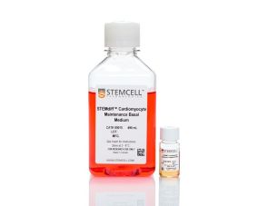
长期维持人类psc来源的心肌细胞的培养基
用于分离hpsc衍生的心肌细胞
解冻和培养hpsc来源的心肌细胞的培养基
一次性使用、免维持的人诱导多能干细胞,冷冻