产品号 #05008_C
无血清和无bpe培养基用于原代人气道上皮细胞的扩增
无血清和无bpe培养基用于原代人气道上皮细胞的扩增
Enzymatic cell dissociation reagent
Cell culture supplement
Hanks' Balanced Salt Solution (HBSS) without calcium and magnesium
Dulbecco’s phosphate-buffered saline without calcium and magnesium
PneumaCult™- ex是一种明确的,不含血清和bpe的细胞培养基,支持人类气道上皮细胞的快速扩增。
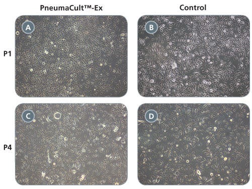
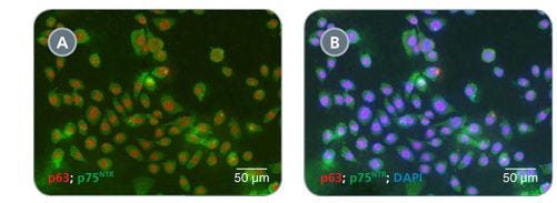
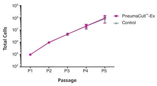
在PneumaCult™-Ex中培养的原代气道上皮细胞在至少3代内迅速扩增,同时保持鹅卵石状形态和基底细胞标记物p63和p75的均匀表达NTR. 此外,在PneumaCult™-Ex中培养的细胞在气液界面中培养时可以分化成假层状粘膜纤毛上皮。
一起,PneumaCult™-Ex和PneumaCult™-ALI构成了一个完全集成的无bpe培养系统,用于体外人体气道建模。这一强大而明确的系统是基础呼吸研究、毒性研究和药物开发的宝贵工具。
了解如何培养人类气道上皮细胞在我们的ALI按需肺课程或浏览我们的常见问题(FAQs)关于使用PneumaCult™的ALI培养工作流程。
Subtype
Specialized Media
Cell Type
Airway Cells
Species
Human
Application
Cell Culture, Expansion, Maintenance
Brand
PneumaCult
Area of Interest
Epithelial Cell Biology
Formulation Category
Serum-Free
Find supporting information and directions for use in the Product Information Sheet or explore additional protocols below.
This product is designed for use in the following research area(s) as part of the highlighted workflow stage(s). Explore these workflows to learn more about the other products we offer to support each research area.
| Species | Human |
|---|---|
| Formulation Category | Serum-Free |
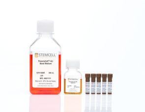
在气液界面培养的人气道上皮细胞的无血清和无bpe培养基
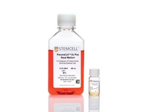
无血清和无bpe培养基用于原代人气道上皮细胞的扩增
哺乳动物活细胞计数试剂