产品号 #05513_C
用于培养小鼠间充质干细胞和骨髓间充质干细胞
用于培养小鼠间充质干细胞和骨髓间充质干细胞
Reagent for manual counting of nucleated mammalian cells
Cell culture supplement
Enzymatic cell dissociation reagent
Cell separation buffer
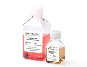
MesenCult™扩增试剂盒(小鼠)是标准的小鼠间充质间质细胞(MSCs)培养;也称为间充质干细胞)和小鼠胚胎成纤维细胞(mef)。该试剂盒包括MesenCult™基础培养基(小鼠),MesenCult™10X补充(小鼠)和MesenPure™。MesenCult™扩增培养基已经过优化,可用于小鼠MSCs和mef的体外衍生和扩增,以及集落形成单位-成纤维细胞(CFU-F)的检测。该试剂盒使用小鼠菌株C57BL/6的细胞进行优化。
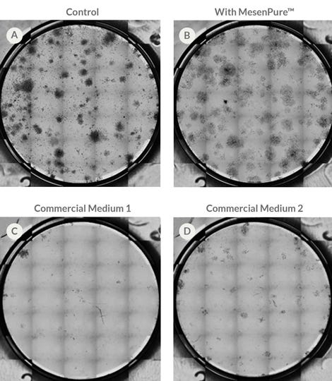
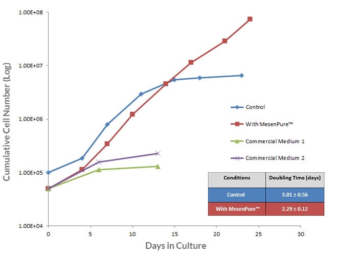
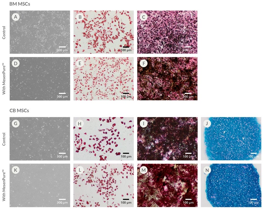
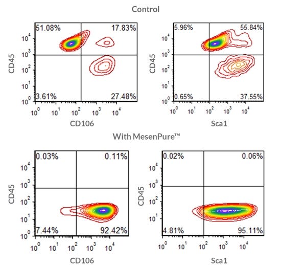
为了促进MSCs和mef在细胞培养过程中的富集,无需连续传代和频繁的培养基变化,只需在使用前添加MesenPure™以完成MesenCult™扩增培养基。虽然不是必需的,但强烈建议添加MesenPure™,因为与单独使用完整的mesenult™扩增培养基相比,所得到的MSC和MEF培养物更均匀,并且表现出更强大的增殖、分化和集落形成。
注意:MesenCult™膨胀介质必须补充l -谷氨酰胺(目录#07100)。
Subtype
Specialized Media
Cell Type
Mesenchymal Stem and Progenitor Cells, Mouse Embryonic Fibroblasts
Species
Mouse
Application
Cell Culture, Colony Assay, Expansion
Brand
MesenCult
Area of Interest
Stem Cell Biology
Find supporting information and directions for use in the Product Information Sheet or explore additional protocols below.
This product is designed for use in the following research area(s) as part of the highlighted workflow stage(s). Explore these workflows to learn more about the other products we offer to support each research area.
| Species | Mouse |
|---|
小鼠间充质干细胞和胚胎成纤维细胞分化成成骨细胞的完全培养基
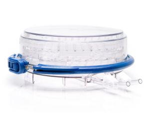
培养室为组织培养提供缺氧环境
Dulbecco的磷酸盐缓冲盐水,不含钙和镁
用于细胞分离和细胞培养的无菌聚丙烯锥形管