EasySep™小鼠MDSC(CD11b+Gr1+)分选试剂盒

免疫磁珠负选未标记的小鼠MDSC(CD11b+Gr1+)细胞

产品号 #19867

免疫磁珠分选未标记的髓源性抑制细胞(CD11b+Gr1+)

产品优势

  • 快速、易于操作,且无需分离柱
  • 纯度高达96%
  • 获得未标记的活细胞

产品组分包括

  • EasySep™小鼠MDSC(CD11b+Gr1+)分选试剂盒(产品号#19867)
    • EasySep™小鼠MDSC(CD11b+Gr1+)分选抗体混合物,0.5mL
    • EasySep™ Streptavidin RapidSpheres™ 50001磁珠,1.0mL
    • EasySep™小鼠FcR阻断剂,2 x 0.2 mL

概述

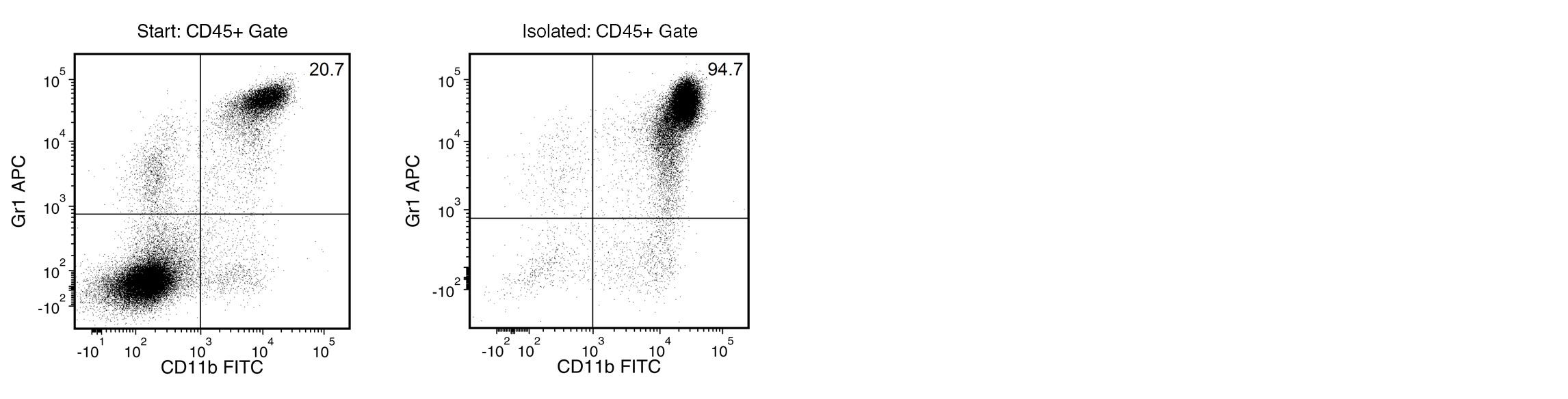
使用EasySep™小鼠MDSC(CD11b+Gr1+)分选试剂盒,通过免疫磁珠负选,可轻松高效地从小鼠脾细胞、骨髓或外周血样本中分离高纯度髓源性抑制细胞(MDSCs)。EasySep™技术结合单克隆抗体的特异性和无需分离柱的简便磁分选系统,已在发表的研究中广泛应用超过20年。
通过EasySep™负选方案,非目标细胞会被抗体复合物与磁珠标记。通过EasySep™磁将被磁珠
标记的细胞与未被标记的目的细胞分离,接着简单地将目的细胞倾倒或吸取至一个新的分离管中。整个磁分选过程仅需18分钟,分离后的MDSCs可立即用于流式细胞术、培养或基于细胞的检测等下游应用。
了解更多关于免疫磁珠EasySep™技术的工作原理。探索更多优化您的实验流程产品,包括培养基、添加剂、抗体等。

MAGNET COMPATIBILITY
• EasySep™ Magnet (Catalog #18000)
• “The Big Easy” EasySep™ Magnet (Catalog #18001)
• EasyEights™ EasySep™ Magnet (Catalog #18103)
 
SUBTYPE
Cell Isolation Kits
 
CELL TYPE
Granulocytes and Subsets, Monocytes, Myeloid Cells
 
SPECIES
Mouse
 
SAMPLE SOURCE
Bone Marrow, Other, Whole Blood
 
SELECTION METHOD
Negative
 
APPLICATION
Cell Isolation
 
BRAND
EasySep
 
AREA OF INTEREST
Immunology

实验数据

Typical EasySep™ Mouse MDSC (CD11b+Gr1+) Cell Isolation Profile from 4T1 Tumor-Bearing BALB/c Mouse Splenocytes

Figure 1. Typical EasySep™ Mouse MDSC (CD11b+Gr1+) Cell Isolation Profile from 4T1 Tumor-Bearing BALB/c Mouse Splenocytes

Starting with 4T1 tumor-bearing BALB/c mouse splenocytes, the MDSC content (CD11b+Gr1+) of the isolated fraction is typically 94.3 ± 2.1% (mean ± SD) using the purple EasySep™ Magnet. In the above example, the purities of the start and final isolated fractions are 20.7% and 94.7%, respectively.

Typical EasySep™ Mouse MDSC (CD11b+Gr1+) Cell Isolation Profile from Naïve C57BL/6 Mouse Splenocytes

Figure 2. Typical EasySep™ Mouse MDSC (CD11b+Gr1+) Cell Isolation Profile from Naïve C57BL/6 Mouse Splenocytes

Starting with naïve C57BL/6 mouse splenocytes, the CD11b+Gr1+ cell content of the isolated fraction is typically 86 ± 4.6% (mean ± SD) using the purple EasySep™ Magnet. In the above example, the purities of the start and final isolated fractions are 3.0% and 86.8%, respectively.

产品说明书及文档

Find supporting information and directions for use in the Product Information Sheet or explore additional protocols below.

Document Type
Product Name
Catalog #
Lot #
Language
Catalog #
19867
Lot #
All
Language
English
Document Type
Safety Data Sheet 1
Catalog #
19867
Lot #
All
Language
English
Document Type
Safety Data Sheet 2
Catalog #
19867
Lot #
All
Language
English
Document Type
Safety Data Sheet 3
Catalog #
19867
Lot #
All
Language
English

应用领域

This product is designed for use in the following research area(s) as part of the highlighted workflow stage(s). Explore these workflows to learn more about the other products we offer to support each research area.

相关材料与文献

Educational Materials (7)

Publications (4)

METTL3 Inhibits Antitumor Immunity by Targeting m6A-BHLHE41-CXCL1/CXCR2 Axis to Promote Colorectal Cancer. H. Chen et al. Gastroenterology 2022 oct

Abstract

BACKGROUND & AIMS N6-Methyladenosine (m6A) is the most prevalent RNA modification and recognized as an important epitranscriptomic mechanism in colorectal cancer (CRC). We aimed to exploit whether and how tumor-intrinsic m6A modification driven by methyltransferase like 3 (METTL3) can dictate the immune landscape of CRC. METHODS Mettl3 knockout mice, CD34+ humanized mice, and different syngeneic mice models were used. Immune cell composition and cytokine level were analyzed by flow cytometry and Cytokine 23-Plex immunoassay, respectively. M6A sequencing and RNA sequencing were performed to identify downstream targets and pathways of METTL3. Human CRC specimens (n = 176) were used to evaluate correlation between METTL3 expression and myeloid-derived suppressor cell (MDSC) infiltration. RESULTS We demonstrated that silencing of METTL3 in CRC cells reduced MDSC accumulation to sustain activation and proliferation of CD4+ and CD8+ T cells, and eventually suppressed CRC in ApcMin/+Mettl3+/- mice, CD34+ humanized mice, and syngeneic mice models. Mechanistically, METTL3 activated the m6A-BHLHE41-CXCL1 axis by analysis of m6A sequencing, RNA sequencing, and cytokine arrays. METTL3 promoted BHLHE41 expression in an m6A-dependent manner, which subsequently induced CXCL1 transcription to enhance MDSC migration in vitro. However, the effect was negligible on BHLHE41 depletion, CXCL1 protein or CXCR2 inhibitor SB265610 administration, inferring that METTL3 promotes MDSC migration via BHLHE41-CXCL1/CXCR2. Consistently, depletion of MDSCs by anti-Gr1 antibody or SB265610 blocked the tumor-promoting effect of METTL3 in vivo. Importantly, targeting METTL3 by METTL3-single guide RNA or specific inhibitor potentiated the effect of anti-programmed cell death protein 1 (anti-PD1) treatment. CONCLUSIONS Our study identifies METTL3 as a potential therapeutic target for CRC immunotherapy whose inhibition reverses immune suppression through the m6A-BHLHE41-CXCL1 axis. METTL3 inhibition plus anti-PD1 treatment shows promising antitumor efficacy against CRC.
STAT3 inhibitor Napabucasin abrogates MDSC immunosuppressive capacity and prolongs survival of melanoma-bearing mice. R. Bitsch et al. Journal for immunotherapy of cancer 2022 mar

Abstract

BACKGROUND Myeloid-derived suppressor cells (MDSCs) represent a negative prognostic factor in malignant melanoma. These cells are generated under chronic inflammatory conditions typical of cancer. The transcription factor signal transducer and activator of transcription 3 (STAT3) orchestrates MDSC accumulation and acquisition of immunosuppressive properties. Here we studied STAT3 inhibition by Napabucasin as a way to block MDSC accumulation and activity and its potential to treat malignant melanoma. METHODS In vitro generated murine MDSC and primary MDSC from melanoma-bearing mice were used to investigate the effects of Napabucasin on MDSC in vitro. The RET transgenic mouse model of malignant melanoma was used to examine Napabucasin therapy efficiency and its underlying mechanisms in vivo. Furthermore, STAT3 activation and its correlation with survival were explored in MDSC from 19 patients with malignant melanoma and human in vitro generated monocytic myeloid-derived suppressor cell (M-MDSC) were used to evaluate the effects of Napabucasin. RESULTS Napabucasin was able to abrogate the capacity of murine MDSC to suppress CD8+ T-cell proliferation. The STAT3 inhibitor induced apoptosis in murine MDSC, significantly increased expression of molecules associated with antigen processing and presentation, as well as slightly decreased expression of immunosuppressive factors on these cells. RET transgenic mice treated with Napabucasin showed prolonged survival accompanied by a strong accumulation of tumor-infiltrating antigen-presenting cells and activation of CD8+ and CD4+ T cells. Interestingly, patients with malignant melanoma with high expression of activated STAT3 in circulating M-MDSC showed significantly worse progression-free survival (PFS) than patients with low levels of activated STAT3. In addition, Napabucasin was able to abrogate suppressive capacity of human in vitro generated M-MDSC. CONCLUSION Our findings demonstrate that STAT3 inhibitor Napabucasin completely abrogated the immunosuppressive capacity of murine MDSC and human M-MDSC and improved melanoma-bearing mouse survival. Moreover, patients with malignant melanoma with high expression levels of activated STAT3 in M-MDSC displayed shorter PFS, indicating its role as a promising therapeutic target in patients with malignant melanoma and a predictive marker for their clinical outcome.
Isolation of human and mouse myeloid-derived suppressor cells for metabolic analysis. J. E. Choi et al. STAR protocols 2022 jun

Abstract

Metabolic reprogramming is associated with myeloid-derived suppressor cell (MDSC) immunosuppressive function. Here, we outline the process for acquiring MDSCs from human and murine sources for subsequent analysis of fatty acid oxidation, oxidative phosphorylation, and glycolysis using the Seahorse XFe 96 Analyzer. Murine MDSCs can be isolated directly from tumor-bearing mice or derived through IL-6 and GM-CSF culture of bone marrow cells from non-tumor-bearing mice. To generate human MDSCs, peripheral blood mononuclear cells (PBMCs) can be cultured with IL-6 and GM-CSF. For complete details on the use and execution of this protocol, please refer to Mohammadpour et al. (2021).
PRODUCTS ARE FOR RESEARCH USE ONLY AND NOT INTENDED FOR HUMAN OR ANIMAL DIAGNOSTIC OR THERAPEUTIC USES UNLESS OTHERWISE STATED. FOR ADDITIONAL INFORMATION ON QUALITY AT STEMCELL, REFER TO WWW.STEMCELL.COM/COMPLIANCE.
版权 © 2025 STEMCELL Technologies 技术有限公司。保留所有权利。