产品号 #03804_C
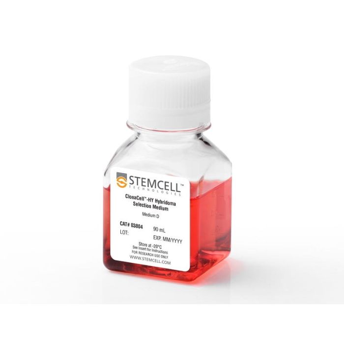
半固体甲基纤维素基杂交瘤筛选和克隆培养基,含HAT(含血清)
通过使用甲基纤维素为基础的培养基一步完成杂交瘤的选择和克隆,节省了研究时间和资源。使用ClonaCell™-HY Medium D容易分离单克隆,单克隆概率高,用于进一步筛选和扩增。
杂交瘤克隆通常具有广泛的生长速率和生产力;当使用ClonaCell™-HY Medium D进行克隆时,它们形成可见且离散的单克隆菌落,防止因过度生长而丢失稀有和高产的克隆。通过人工或机器人的方法,可以很容易地从半固体培养基中挑选杂交瘤菌落,并分散到液体培养基中进行筛选和扩增。
ClonaCell™-HY Medium D是一种半固体培养基,含有血清和选择性试剂次黄嘌呤、氨基蝶呤和胸腺嘧啶(HAT)。它已被证实用于发展小鼠和大鼠杂交瘤。也有报道称,它与使用来自多种宿主动物(包括人类、小鼠、大鼠和仓鼠)的淋巴细胞生产和克隆杂交瘤兼容(例如Wilson JR等)。抗病毒研究,2016)。
为什么使用半固态克隆:
•形成物理上分离的,离散的菌落,可以很容易地分离。
•与散装液体培养相比,在半固体培养基中同时选择和克隆容易分离稀有和高产克隆。
•与限制稀释的选择和克隆相比,用更少的时间和资源分离单克隆细胞系。
找到更多的产品,高效的杂交瘤克隆和一代,在我们的ClonaCell™品牌页面。
Contains
• DMEM
• Methylcellulose
• Serum
• Hypoxanthine, aminopterin, and thymidine (HAT)
• Gentamicin
• 2-Mercaptoethanol
• Phenol red
• L-Glutamine and other supplements
• Other ingredients
Subtype
Semi-Solid Media, Specialized Media
Cell Type
Hybridomas
Species
Human, Mouse, Other, Rat
Application
Cell Culture, Semi-Solid Cloning
Brand
ClonaCell
Area of Interest
Antibody Development, Cancer, Cell Line Development, Drug Discovery and Toxicity Testing, Hybridoma Generation
Formulation Category
Methylcellulose-Based
Find supporting information and directions for use in the Product Information Sheet or explore additional protocols below.
This product is designed for use in the following research area(s) as part of the highlighted workflow stage(s). Explore these workflows to learn more about the other products we offer to support each research area.
| Species | Human, Mouse, Other, Rat |
|---|---|
| Contains | • DMEM • Methylcellulose • Serum • Hypoxanthine, aminopterin, and thymidine (HAT) • Gentamicin • 2-Mercaptoethanol • Phenol red • L-Glutamine and other supplements • Other ingredients |
| Formulation Category | Methylcellulose-Based |
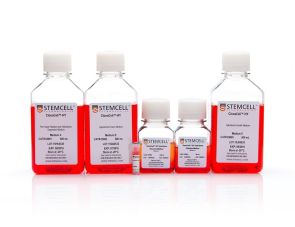
完整的杂交瘤生成试剂盒
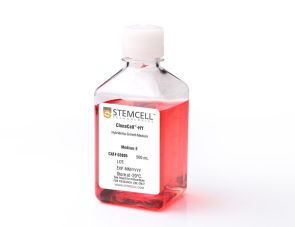
次黄嘌呤胸腺嘧啶杂交瘤生长培养基(含血清)
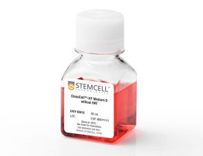
半固体甲基纤维素基杂交瘤筛选和克隆培养基,不含HAT(含血清)