产品号 #60002_C
抗小鼠CD11c的仓鼠(亚美尼亚)单克隆IgG抗体
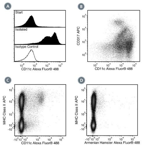
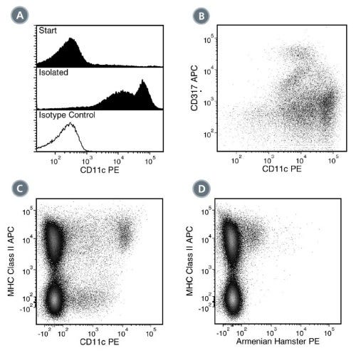
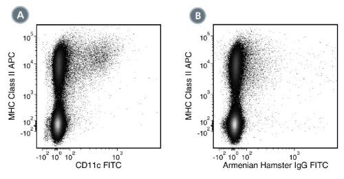
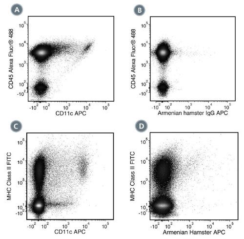
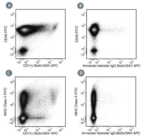
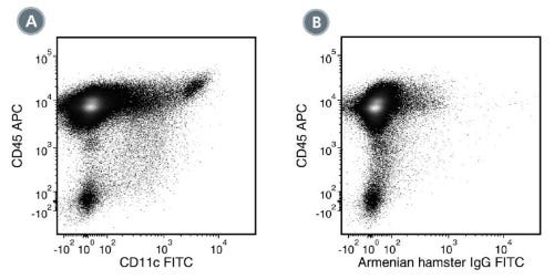
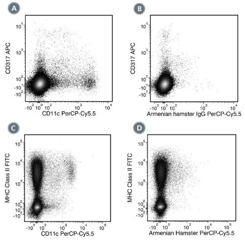
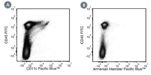
N418抗体可靶向CD11c(αX整合素)。CD11c是一种150 kDa的1型跨膜糖蛋白,其通过与CD18(β2整合素)非共价结合形成异二聚体细胞表面黏附受体。CD11c/CD18受体通过结合iC3b、纤维蛋白原和CD54等配体,参与多种免疫应答过程,包括细胞迁移、刺激单核细胞和巨噬细胞产生细胞因子、T细胞扩增、白细胞募集和吞噬作用。在小鼠中,CD11c在树突状细胞、巨噬细胞、单核细胞、粒细胞、NK细胞和部分T细胞上表达。
N418抗体克隆已通过验证,适用于EasySep™试剂盒(包括EasySep™小鼠CD11c正选试剂盒II,产品号 #18780)分离的细胞纯度检测。
亚型
一抗
靶抗原
CD11c
别名
alphaX整合素,CR4,整合素alphaX链,p150
活性物种
小鼠
偶联
Biotin 或 生物素,FITC,PE,未偶联的
宿主物种
Hamster
细胞类型
树突状细胞(DCs)
种属
小鼠
应用
CyTOF,流式细胞术,免疫细胞化学,免疫荧光,免疫组化,免疫沉淀
研究领域
免疫
克隆
N418
基因编号
16411
同种型
IgG
Find supporting information and directions for use in the Product Information Sheet or explore additional protocols below.
This product is designed for use in the following research area(s) as part of the highlighted workflow stage(s). Explore these workflows to learn more about the other products we offer to support each research area.
| Species | Mouse |
|---|---|
| Clone | N418 |
| Gene Id | 16411 |
| Alternative Names | alphaX integrin, CR4, integrin alphaX chain, p150 |
| Isotype | IgG |
免疫磁珠正选小鼠CD11c+细胞
免疫磁珠负选试剂盒