产品号 #100-0852_C
适用于细胞培养中F-肌动蛋白检测的荧光染料偶联物
鬼笔环肽是一种七肽,来源于有毒真菌毒蝇伞(Amanita phalloides)。鬼笔环肽可高特异性地结合细胞内的F-肌动蛋白(F-actin)丝,并可防止其去聚合。荧光标记的鬼笔环肽可用于对经甲醛固定并通透处理后的细胞中F-actin的标记、识别与定量分析。与大多数动植物物种的肌动蛋白抗体相比,荧光鬼笔环肽表现出更低的非特异性结合,从而在成像过程中提供较低背景信号和更高图像对比度。
细胞类型
神经细胞,PSC衍生,神经元,多能干细胞
应用
鉴定
研究领域
癌症,神经科学,干细胞生物学
CAS 编号
67-68-5
分子量
鬼笔环肽,iFluor™ 488*: 1399.49 克/摩尔; 鬼笔环肽,iFluor™ 555*: 1468.53 克/摩尔; 鬼笔环肽,iFluor™ 647: 1632.87 克/摩尔
Find supporting information and directions for use in the Product Information Sheet or explore additional protocols below.
This product is designed for use in the following research area(s) as part of the highlighted workflow stage(s). Explore these workflows to learn more about the other products we offer to support each research area.
| Molecular Weight | Phalloidin, iFluor™ 488*: 1399.49 g/mol; Phalloidin, iFluor™ 555*: 1468.53 g/mol; Phalloidin, iFluor™ 647: 1632.87 g/mol |
|---|---|
| Cas Number | 67-68-5 |

提升神经元功能的无血清基础培养基
无血清神经生理基础培养基,改善神经元活细胞成像和功能
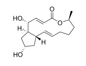
蛋白质运输抑制剂;抑制含sec7的鸟嘌呤交换因子(GEF)