紫杉醇

微管形成抑制剂

产品号 #(选择产品)

产品号 #73312_C

微管形成抑制剂

总览

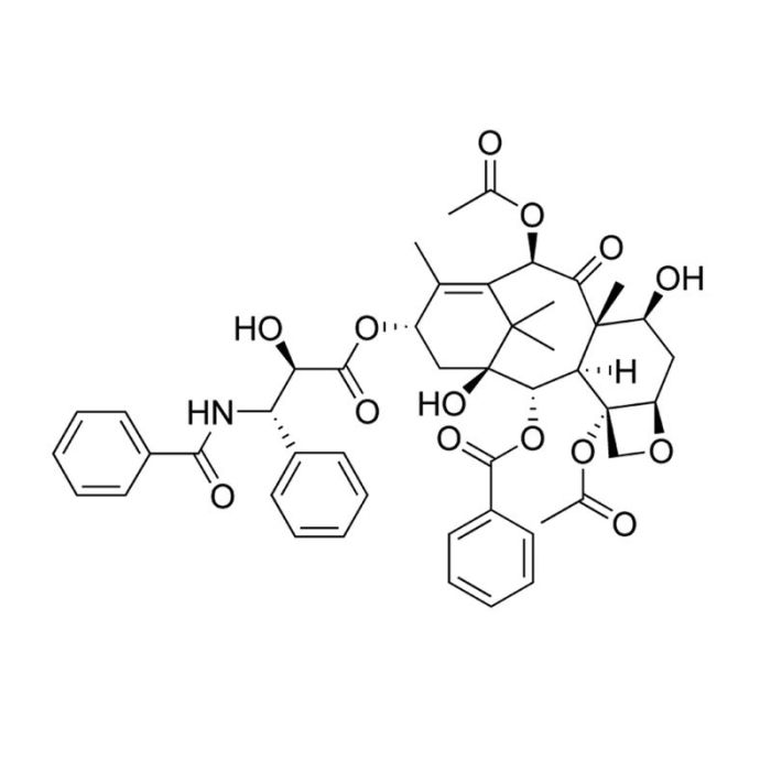
紫杉醇是一种二萜生物碱,最初从太平洋紫杉(Taxus brevifolia)树皮中分离得到。它能结合并稳定微管,阻止其在细胞分裂过程中重组,从而导致细胞周期停滞。紫杉醇具有抗肿瘤特性,并已被用作化疗药物(Rowinsky等人)。许多通路与紫杉醇诱导的细胞凋亡有关,包括c-Jun 氨基末端激酶/应激激活蛋白激酶(JNK/SAPK),p38丝裂原活化蛋白激酶(MAPK)和蛋白激酶A (PKA;Wang等;Reshkin等)。

分化
·通过微管聚合抑制体外神经突的形成和生长(Letourneau和Ressler)。

癌症研究
·抑制多种癌细胞系的肿瘤细胞生长,包括宫颈癌(HeLa)、肺癌(A549)、乳腺癌(MCF-7)、结肠癌(HT-29)、卵巢癌(OVG-1)和胰腺癌(PC-Sh)(Liebmann等)。
·诱导异常多极纺锤体形成,诱导多种人类细胞癌细胞系中细胞周期停滞于前期和 G1 期(Woods等)。
·通过多种机制启动癌细胞凋亡,包括:p53依赖性和非依赖性通路、B细胞CLL/淋巴瘤2(BCL-2)家族成员、细胞周期依赖性蛋白激酶、p38 MAPK、PKA和JNK/SAPK(Wang等;Reshkin等)。
·通过依赖于RAF-1激活的机制诱导MCF7和PC3M人类癌细胞系中的细胞周期蛋白抑制剂p21(Blagosklonny等)。

细胞类型
癌细胞及细胞系,白血病/淋巴瘤细胞,神经元
 
种属
人,小鼠,非人灵长类,其它细胞系,大鼠
 
研究领域
癌症,神经科学
 
CAS 编号
33069-62-4
 
化学式
C₄₇H₅₁NO₁₄
 
纯度
≥98%
 

Protocols and Documentation

Find supporting information and directions for use in the Product Information Sheet or explore additional protocols below.

Document Type
Product Name
Catalog #
Lot #
Language
Product Name
Paclitaxel
Catalog #
73312, 73314
Lot #
All
Language
English
Document Type
Safety Data Sheet
Product Name
Paclitaxel
Catalog #
73312, 73314
Lot #
All
Language
English

Applications

This product is designed for use in the following research area(s) as part of the highlighted workflow stage(s). Explore these workflows to learn more about the other products we offer to support each research area.

Resources and Publications

Educational Materials (2)

Publications (7)

Paclitaxel induces apoptosis via protein kinase A- and p38 mitogen-activated protein-dependent inhibition of the Na+/H+ exchanger (NHE) NHE isoform 1 in human breast cancer cells. Reshkin SJ et al. Clinical cancer research : an official journal of the American Association for Cancer Research 2003

Abstract

PURPOSE: The molecular signal components essential to paclitaxel-dependent apoptosis in breast cancers are potential targets for combined therapy. However, the signal mechanisms underlying paclitaxel action still need to be better defined. EXPERIMENTAL DESIGN: In a breast cancer cell line, pharmacological agents and transient transfection with dominant interfering and constitutive active mutants were used to identify the signal transduction module involved in the regulation of paclitaxel-induced apoptosis and to evaluate its potential as a therapeutic target. RESULTS: In MDA-MB-435 cells, paclitaxel treatment stimulated the activity of both protein kinase A and p38, and inhibited the activity of the Na(+)/H(+) exchanger isoform 1 (NHE1) with similar IC(50) concentrations as for its activation of apoptosis. Activation and inhibition experiments demonstrated that protein kinase A and p38 participate sequentially upstream of the NHE1 in regulating the paclitaxel-induced apoptotic pathway. Importantly, concurrent specific inhibition of the NHE1 with paclitaxel treatment resulted in a synergistic induction of apoptosis and a reduction in the paclitaxel IC(50) for apoptosis. This sensitization of paclitaxel apoptotic action by specific inhibition of NHE1 was verified in breast cancer cell lines with different paclitaxel sensitivity. CONCLUSIONS: We have, for the first time, identified NHE1 as an essential component of paclitaxel-induced apoptosis in breast cancer cells and, importantly, identified that simultaneous inhibition of the NHE1 results in a synergistic potentiation of low-dose paclitaxel apoptotic action. As specific NHE1 inhibitors have finished Phase II/Phase III clinical trials for myocardial protection, there is the possibility for a rapid biological translation of this novel therapeutic strategy to a clinical setting.
Paclitaxel-induced cell death: where the cell cycle and apoptosis come together. Wang TH et al. Cancer 2000

Abstract

BACKGROUND: Compelling evidence indicates that paclitaxel kills cancer cells through the induction of apoptosis. Paclitaxel binds microtubules and causes kinetic suppression (stabilization) of microtubule dynamics. The consequent arrest of the cell cycle at mitotic phase has been considered to be the cause of paclitaxel-induced cytotoxicity. However, the biochemical events, downstream from paclitaxel's binding to microtubules, that lead to apoptosis are not well understood. METHODS: The authors examined recent scientific literature about the mechanisms by which paclitaxel exerts cytotoxicity. RESULTS: In addition to an arrest of the cell cycle at the mitotic phase in paclitaxel-treated cells, recent discoveries of activation of signaling molecules by paclitaxel and paclitaxel-induced transcriptional activation of various genes indicate that paclitaxel initiates apoptosis through multiple mechanisms. The checkpoint of mitotic spindle assembly, aberrant activation of cyclin-dependent kinases, and the c-Jun N-terminal kinase/stress-activated protein kinase (JNK/SAPK) are shown to be involved in paclitaxel-induced apoptosis. Consistent with observations that microtubules of different status (e.g., cytoskeletal microtubules vs. mitotic spindles) have different sensitivity to paclitaxel, the concentration of paclitaxel appears to be the major determinant of its apoptogenic mechanisms. CONCLUSIONS: Advances in research of the cell cycle and apoptosis have extended our understanding of the mechanisms of paclitaxel-induced cell death. Further elucidation of resistance and enhancement of paclitaxel-induced apoptosis should expedite the development of better paclitaxel-based regimens for cancer therapy.
Taxol-induced mitotic block triggers rapid onset of a p53-independent apoptotic pathway. Woods CM et al. Molecular medicine (Cambridge, Mass.) 1995

Abstract

BACKGROUND: At therapeutic concentrations, the antineoplastic agent taxol selectively perturbs mitotic spindle microtubules. Taxol has recently been shown to induce apoptosis, similar to the mechanism of cell death induced by other antineoplastic agents. However, taxol has shown efficacy against drug-refractory cancers, raising the possibility that this pharmacological agent may trigger an alternative apoptotic pathway. MATERIALS AND METHODS: The kinetics and IC50 of mitotic (M) block, aberrant mitosis, and cytotoxicity following taxol treatment were analyzed in human cell lines as well as normal mouse embryo fibroblasts (MEFs) and MEFs derived from p53-null mice. Apoptosis was followed by DNA gel electrophoresis and by in situ DNA end-labeling (TUNEL). RESULTS: Taxol induced two forms of cell cycle arrest: either directly in early M at prophase or, for those cells progressing through aberrant mitosis, arrest in G1 as multimininucleated cells. TUNEL labeling revealed that DNA nicking occurred within 30 min of the arrest in prophase. In contrast, G1-arrested, multimininucleated cells became TUNEL positive only after several days. In the subset of cells that became blocked directly in prophase, both wt p53-expressing and p53-null MEFs responded similarly to taxol, showing rapid onset of DNA nicking and apoptosis. However, p53-null MEFs progressing through aberrant mitosis failed to arrest in the subsequent G1 phase or to become TUNEL positive, and remained viable. CONCLUSIONS: Taxol induces two forms of cell cycle arrest, which in turn induce two independent apoptotic pathways. Arrest in prophase induces rapid onset of a p53-independent pathway, whereas G1-block and the resulting slow (3-5 days) apoptotic pathway are p53 dependent.

更多信息

更多信息
Species Human, Mouse, Non-Human Primate, Other, Rat
Cas Number 33069-62-4
Chemical Formula C₄₇H₅₁NO₁₄
Purity ≥ 98%
PRODUCTS ARE FOR RESEARCH USE ONLY AND NOT INTENDED FOR HUMAN OR ANIMAL DIAGNOSTIC OR THERAPEUTIC USES UNLESS OTHERWISE STATED. FOR ADDITIONAL INFORMATION ON QUALITY AT STEMCELL, REFER TO WWW.STEMCELL.COM/COMPLIANCE. Safety Statement: CA WARNING: This product can expose you to Paclitaxel which is known to the State of California to cause birth defects or other reproductive harm. For more information go to www.P65Warnings.ca.gov
版权 © 2025 STEMCELL Technologies 技术有限公司。保留所有权利。