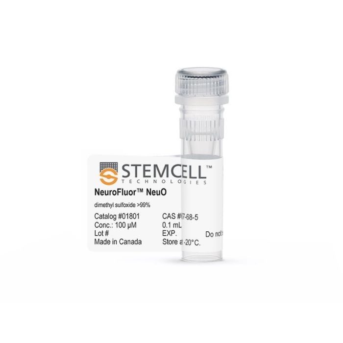
产品号 #01801_C
用于检测活神经元的透膜荧光探针
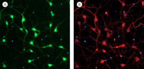
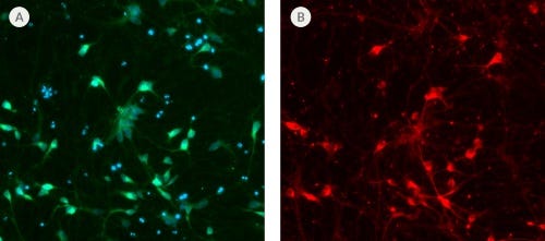
NeuroFluor™NeuO是一种膜渗透荧光探针,可在其他脑细胞存在的情况下选择性标记原代和多能干细胞来源的神经元(J C Er等)。新化学,2015)。用NeuroFluor™NeuO标记的细胞可以使用荧光成像可视化。用这种探针标记是非永久性的;它可以洗掉,为下游应用提供未标记的活细胞。有关其他信息,请参见参考资料。
Contains
• 100 µM NeuO (CAS Number: 1616355-50-0, 1668597-38-3)
• DMSO
Cell Type
Neural Cells, PSC-Derived, Neurons
Species
Human, Mouse, Rat
Application
Characterization, Phenotyping
Brand
NeuroFluor
Area of Interest
Disease Modeling, Neuroscience, Stem Cell Biology
Find supporting information and directions for use in the Product Information Sheet or explore additional protocols below.
This product is designed for use in the following research area(s) as part of the highlighted workflow stage(s). Explore these workflows to learn more about the other products we offer to support each research area.
| Species | Human, Mouse, Rat |
|---|---|
| Contains | • 100 µM NeuO (CAS Number: 1616355-50-0, 1668597-38-3) • DMSO |
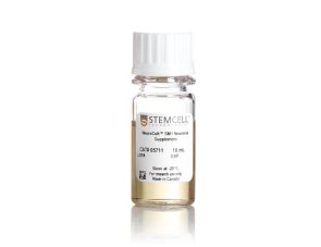
无血清神经添加物(50X)
提升神经元功能的无血清基础培养基